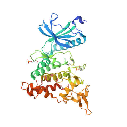
4NCT

Human DYRK1A in complex with PKC412


Experimental Data Snapshot

  • Method: X-RAY DIFFRACTION
  • Resolution: 2.60 Å
  • R-Value Free: 0.249 
  • R-Value Work: 0.208 
  • R-Value Observed: 0.210 

wwPDB Validation   3D Report Full Report


Ligand Structure Quality Assessment 


This is version 1.3 of the entry. See complete history


Literature

The structure of a dual-specificity tyrosine phosphorylation-regulated kinase 1A-PKC412 complex reveals disulfide-bridge formation with the anomalous catalytic loop HRD(HCD) cysteine.

Alexeeva, M.Aberg, E.Engh, R.A.Rothweiler, U.

(2015) Acta Crystallogr D Biol Crystallogr 71: 1207-1215

  • DOI: https://doi.org/10.1107/S1399004715005106
  • Primary Citation of Related Structures:  
    4NCT

  • PubMed Abstract: 

    Dual-specificity tyrosine phosphorylation-regulated kinase 1A (DYRK1A) is a protein kinase associated with neuronal development and brain physiology. The DYRK kinases are very unusual with respect to the sequence of the catalytic loop, in which the otherwise highly conserved arginine of the HRD motif is replaced by a cysteine. This replacement, along with the proximity of a potential disulfide-bridge partner from the activation segment, implies a potential for redox control of DYRK family activities. Here, the crystal structure of DYRK1A bound to PKC412 is reported, showing the formation of the disulfide bridge and associated conformational changes of the activation loop. The DYRK kinases represent emerging drug targets for several neurological diseases as well as cancer. The observation of distinct activation states may impact strategies for drug targeting. In addition, the characterization of PKC412 binding offers new insights for DYRK inhibitor discovery.


  • Organizational Affiliation

    Department of Chemistry, The Norwegian Structural Biology Centre, The Arctic University of Norway, 9037 Tromsø, Norway.


Macromolecules
Find similar proteins by:  (by identity cutoff)  |  3D Structure
Entity ID: 1
MoleculeChains Sequence LengthOrganismDetailsImage
Dual specificity tyrosine-phosphorylation-regulated kinase 1A
A, B, D
368Homo sapiensMutation(s): 0 
Gene Names: DYRK1ADYRKMNBMNBH
EC: 2.7.12.1 (PDB Primary Data), 2.7.11.23 (UniProt)
UniProt & NIH Common Fund Data Resources
Find proteins for Q13627 (Homo sapiens)
Explore Q13627 
Go to UniProtKB:  Q13627
PHAROS:  Q13627
GTEx:  ENSG00000157540 
Entity Groups  
Sequence Clusters30% Identity50% Identity70% Identity90% Identity95% Identity100% Identity
UniProt GroupQ13627
Sequence Annotations
Expand
  • Reference Sequence
Find similar proteins by:  (by identity cutoff)  |  3D Structure
Entity ID: 2
MoleculeChains Sequence LengthOrganismDetailsImage
Dual specificity tyrosine-phosphorylation-regulated kinase 1A368Homo sapiensMutation(s): 0 
Gene Names: DYRK1ADYRKMNBMNBH
EC: 2.7.12.1 (PDB Primary Data), 2.7.11.23 (UniProt)
UniProt & NIH Common Fund Data Resources
Find proteins for Q13627 (Homo sapiens)
Explore Q13627 
Go to UniProtKB:  Q13627
PHAROS:  Q13627
GTEx:  ENSG00000157540 
Entity Groups  
Sequence Clusters30% Identity50% Identity70% Identity90% Identity95% Identity100% Identity
UniProt GroupQ13627
Sequence Annotations
Expand
  • Reference Sequence
Experimental Data & Validation

Experimental Data

  • Method: X-RAY DIFFRACTION
  • Resolution: 2.60 Å
  • R-Value Free: 0.249 
  • R-Value Work: 0.208 
  • R-Value Observed: 0.210 
  • Space Group: P 21 21 21
Unit Cell:
Length ( Å )Angle ( ˚ )
a = 87.764α = 90
b = 87.895β = 90
c = 229.921γ = 90
Software Package:
Software NamePurpose
MxCuBEdata collection
MOLREPphasing
REFMACrefinement
XDSdata reduction
XDSdata scaling

Structure Validation

View Full Validation Report


Ligand Structure Quality Assessment 


Entry History 

Deposition Data

Revision History  (Full details and data files)

  • Version 1.0: 2015-04-01
    Type: Initial release
  • Version 1.1: 2015-05-20
    Changes: Database references
  • Version 1.2: 2015-07-22
    Changes: Database references
  • Version 1.3: 2017-11-15
    Changes: Refinement description