4XO0

Crystal structure of 5'-CTTATPPTAZZATAAG in a host-guest complex


Experimental Data Snapshot

  • Method: X-RAY DIFFRACTION
  • Resolution: 1.70 Å
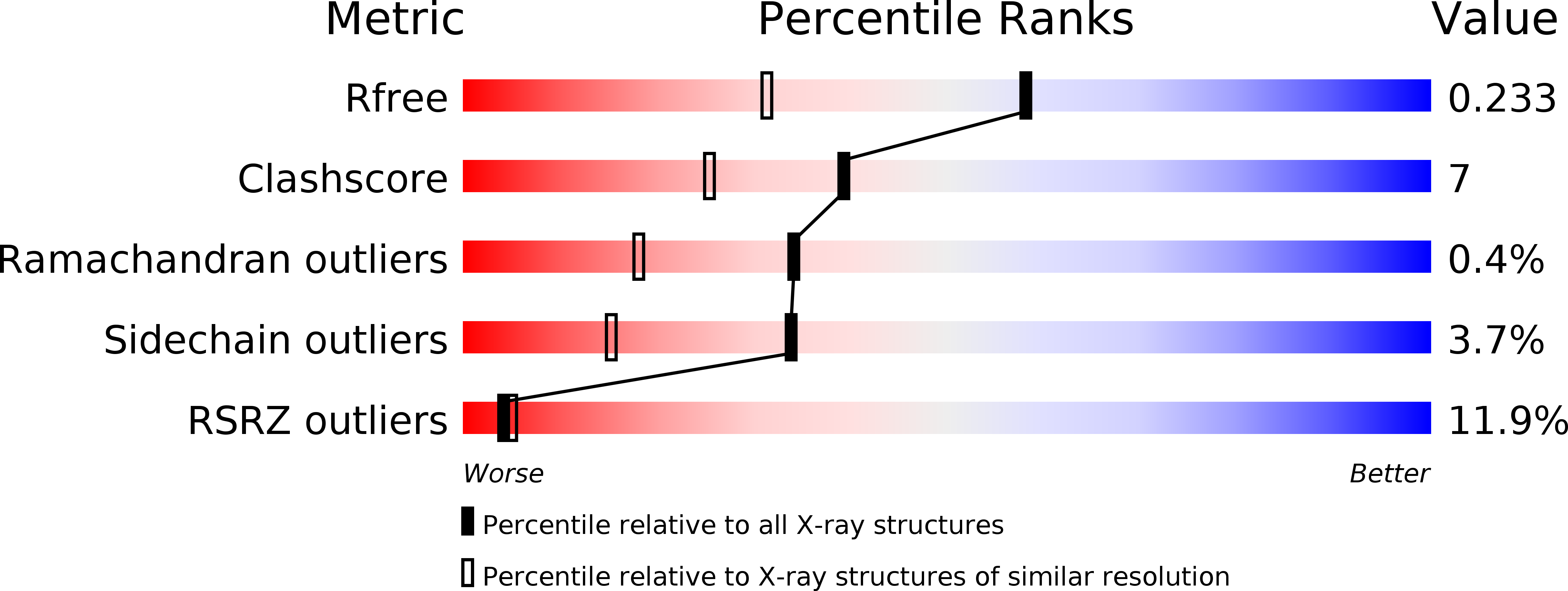
  • R-Value Free: 0.240 
  • R-Value Work: 0.220 
  • R-Value Observed: 0.221 

wwPDB Validation   3D Report Full Report


This is version 1.5 of the entry. See complete history


Literature

Structural basis for a six nucleotide genetic alphabet.

Georgiadis, M.M.Singh, I.Kellett, W.F.Hoshika, S.Benner, S.A.Richards, N.G.

(2015) J Am Chem Soc 137: 6947-6955

  • DOI: https://doi.org/10.1021/jacs.5b03482
  • Primary Citation of Related Structures:  
    4XNO, 4XO0, 4XPC, 4XPE

  • PubMed Abstract: 

    Expanded genetic systems are most likely to work with natural enzymes if the added nucleotides pair with geometries that are similar to those displayed by standard duplex DNA. Here, we present crystal structures of 16-mer duplexes showing this to be the case with two nonstandard nucleobases (Z, 6-amino-5-nitro-2(1H)-pyridone and P, 2-amino-imidazo[1,2-a]-1,3,5-triazin-4(8H)one) that were designed to form a Z:P pair with a standard "edge on" Watson-Crick geometry, but joined by rearranged hydrogen bond donor and acceptor groups. One duplex, with four Z:P pairs, was crystallized with a reverse transcriptase host and adopts primarily a B-form. Another contained six consecutive Z:P pairs; it crystallized without a host in an A-form. In both structures, Z:P pairs fit canonical nucleobase hydrogen-bonding parameters and known DNA helical forms. Unique features include stacking of the nitro group on Z with the adjacent nucleobase ring in the A-form duplex. In both B- and A-duplexes, major groove widths for the Z:P pairs are approximately 1 Å wider than those of comparable G:C pairs, perhaps to accommodate the large nitro group on Z. Otherwise, ZP-rich DNA had many of the same properties as CG-rich DNA, a conclusion supported by circular dichroism studies in solution. The ability of standard duplexes to accommodate multiple and consecutive Z:P pairs is consistent with the ability of natural polymerases to biosynthesize those pairs. This, in turn, implies that the GACTZP synthetic genetic system can explore the entire expanded sequence space that additional nucleotides create, a major step forward in this area of synthetic biology.


Macromolecules

Find similar proteins by:  (by identity cutoff)  |  3D Structure
Entity ID: 1
MoleculeChains Sequence LengthOrganismDetailsImage
reverse transcriptase259Moloney murine leukemia virus isolate ShinnickMutation(s): 0 
Gene Names: gag-pol
EC: 2.7.7.49 (PDB Primary Data), 2.7.7.7 (PDB Primary Data), 3.1.26.4 (PDB Primary Data)
UniProt
Find proteins for P03355 (Moloney murine leukemia virus (isolate Shinnick))
Explore P03355 
Go to UniProtKB:  P03355
Entity Groups  
Sequence Clusters30% Identity50% Identity70% Identity90% Identity95% Identity100% Identity
UniProt GroupP03355
Sequence Annotations
Expand
  • Reference Sequence

Find similar nucleic acids by:  Sequence   |   3D Structure  

Entity ID: 2
MoleculeChains LengthOrganismImage
DNA (5'-D(*CP*TP*TP*AP*TP*(1WA)P*(1WA)P*T)-3')8synthetic construct
Sequence Annotations
Expand
  • Reference Sequence

Find similar nucleic acids by:  Sequence   |   3D Structure  

Entity ID: 3
MoleculeChains LengthOrganismImage
DNA (5'-D(P*AP*(1W5)P*(1W5)P*AP*TP*AP*AP*G)-3')C [auth G]8synthetic construct
Sequence Annotations
Expand
  • Reference Sequence
Experimental Data & Validation

Experimental Data

  • Method: X-RAY DIFFRACTION
  • Resolution: 1.70 Å
  • R-Value Free: 0.240 
  • R-Value Work: 0.220 
  • R-Value Observed: 0.221 
  • Space Group: P 21 21 2
Unit Cell:
Length ( Å )Angle ( ˚ )
a = 54.638α = 90
b = 145.359β = 90
c = 46.878γ = 90
Software Package:
Software NamePurpose
PHENIXrefinement
SCALEPACKdata scaling
PDB_EXTRACTdata extraction
PHASERphasing

Structure Validation

View Full Validation Report


Entry History & Funding Information

Deposition Data


Funding OrganizationLocationGrant Number
National Institutes of Health/National Institute of General Medical Sciences (NIH/NIGMS)United StatesR01GM111386

Revision History  (Full details and data files)

  • Version 1.0: 2015-05-27
    Type: Initial release
  • Version 1.1: 2016-05-18
    Changes: Non-polymer description
  • Version 1.2: 2017-01-11
    Changes: Database references
  • Version 1.3: 2017-09-20
    Changes: Author supporting evidence, Refinement description
  • Version 1.4: 2019-12-25
    Changes: Author supporting evidence
  • Version 1.5: 2024-02-28
    Changes: Data collection, Database references