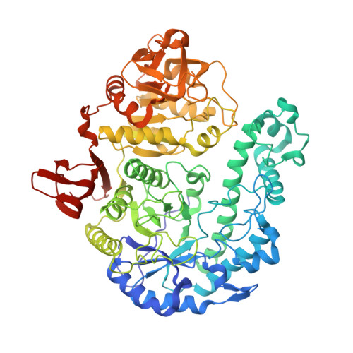
4UNI

beta-(1,6)-galactosidase from Bifidobacterium animalis subsp. lactis Bl-04 in complex with galactose


Experimental Data Snapshot

  • Method: X-RAY DIFFRACTION
  • Resolution: 2.60 Å
  • R-Value Free: 0.234 
  • R-Value Work: 0.175 
  • R-Value Observed: 0.177 

Starting Model: experimental
View more details

wwPDB Validation   3D Report Full Report


Ligand Structure Quality Assessment 


This is version 3.2 of the entry. See complete history


Literature

A beta 1-6/ beta 1-3 galactosidase from Bifidobacterium animalis subsp. lactis Bl-04 gives insight into sub-specificities of beta-galactoside catabolism within Bifidobacterium.

Viborg, A.H.Fredslund, F.Katayama, T.Nielsen, S.K.Svensson, B.Kitaoka, M.Lo Leggio, L.Abou Hachem, M.

(2014) Mol Microbiol 

  • DOI: https://doi.org/10.1111/mmi.12815
  • Primary Citation of Related Structures:  
    4UNI, 4UOQ, 4UOZ

  • PubMed Abstract: 

    The Bifidobacterium genus harbours several health promoting members of the gut microbiota. Bifidobacteria display metabolic specialization by preferentially utilizing dietary or host-derived β-galactosides. This study investigates the biochemistry and structure of a glycoside hydrolase family 42 (GH42) β-galactosidase from the probiotic Bifidobacterium animalis subsp. lactis Bl-04 (BlGal42A). BlGal42A displays a preference for undecorated β1-6 and β1-3 linked galactosides and populates a phylogenetic cluster with close bifidobacterial homologues implicated in the utilization of N-acetyl substituted β1-3 galactosides from human milk and mucin. A long loop containing an invariant tryptophan in GH42, proposed to bind substrate at subsite + 1, is identified here as specificity signature within this clade of bifidobacterial enzymes. Galactose binding at the subsite - 1 of the active site induced conformational changes resulting in an extra polar interaction and the ordering of a flexible loop that narrows the active site. The amino acid sequence of this loop provides an additional specificity signature within this GH42 clade. The phylogenetic relatedness of enzymes targeting β1-6 and β1-3 galactosides likely reflects structural differences between these substrates and β1-4 galactosides, containing an axial galactosidic bond. These data advance our molecular understanding of the evolution of sub-specificities that support metabolic specialization in the gut niche.


  • Organizational Affiliation

    Enzyme and Protein Chemistry, Department of Systems Biology, Technical University of Denmark, DK-2800, Kgs. Lyngby, Denmark.


Macromolecules
Find similar proteins by:  (by identity cutoff)  |  3D Structure
Entity ID: 1
MoleculeChains Sequence LengthOrganismDetailsImage
BETA-GALACTOSIDASE
A, B, C
695Bifidobacterium animalis subsp. lactis Bl-04Mutation(s): 0 
EC: 3.2.1.23
Entity Groups  
Sequence Clusters30% Identity50% Identity70% Identity90% Identity95% Identity100% Identity
Sequence Annotations
Expand
  • Reference Sequence
Small Molecules
Ligands 5 Unique
IDChains Name / Formula / InChI Key2D Diagram3D Interactions
GAL
Query on GAL

Download Ideal Coordinates CCD File 
D [auth A],
K [auth B],
N [auth C]
beta-D-galactopyranose
C6 H12 O6
WQZGKKKJIJFFOK-FPRJBGLDSA-N
PGE
Query on PGE

Download Ideal Coordinates CCD File 
E [auth A]
F [auth A]
G [auth A]
L [auth B]
Q [auth C]
E [auth A],
F [auth A],
G [auth A],
L [auth B],
Q [auth C],
T [auth C]
TRIETHYLENE GLYCOL
C6 H14 O4
ZIBGPFATKBEMQZ-UHFFFAOYSA-N
PEG
Query on PEG

Download Ideal Coordinates CCD File 
H [auth A]
I [auth A]
M [auth B]
P [auth C]
R [auth C]
H [auth A],
I [auth A],
M [auth B],
P [auth C],
R [auth C],
S [auth C]
DI(HYDROXYETHYL)ETHER
C4 H10 O3
MTHSVFCYNBDYFN-UHFFFAOYSA-N
GOL
Query on GOL

Download Ideal Coordinates CCD File 
J [auth A]GLYCEROL
C3 H8 O3
PEDCQBHIVMGVHV-UHFFFAOYSA-N
ZN
Query on ZN

Download Ideal Coordinates CCD File 
O [auth C]ZINC ION
Zn
PTFCDOFLOPIGGS-UHFFFAOYSA-N
Experimental Data & Validation

Experimental Data

  • Method: X-RAY DIFFRACTION
  • Resolution: 2.60 Å
  • R-Value Free: 0.234 
  • R-Value Work: 0.175 
  • R-Value Observed: 0.177 
  • Space Group: C 2 2 21
Unit Cell:
Length ( Å )Angle ( ˚ )
a = 139.52α = 90
b = 199.44β = 90
c = 217.75γ = 90
Software Package:
Software NamePurpose
PHENIXrefinement
XDSdata reduction
XSCALEdata scaling
PHASERphasing

Structure Validation

View Full Validation Report


Ligand Structure Quality Assessment 


Entry History 

Deposition Data

Revision History  (Full details and data files)

  • Version 1.0: 2014-10-15
    Type: Initial release
  • Version 2.0: 2018-01-17
    Changes: Atomic model, Data collection
  • Version 3.0: 2019-02-27
    Changes: Atomic model, Data collection, Database references
  • Version 3.1: 2020-07-29
    Type: Remediation
    Reason: Carbohydrate remediation
    Changes: Data collection, Derived calculations, Other, Structure summary
  • Version 3.2: 2024-01-10
    Changes: Data collection, Database references, Refinement description, Structure summary