4U9D

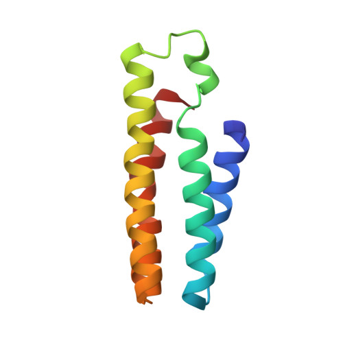
Crystal Structure of the Zn-directed tetramer of the engineered cyt cb562 variant, AB3


Experimental Data Snapshot

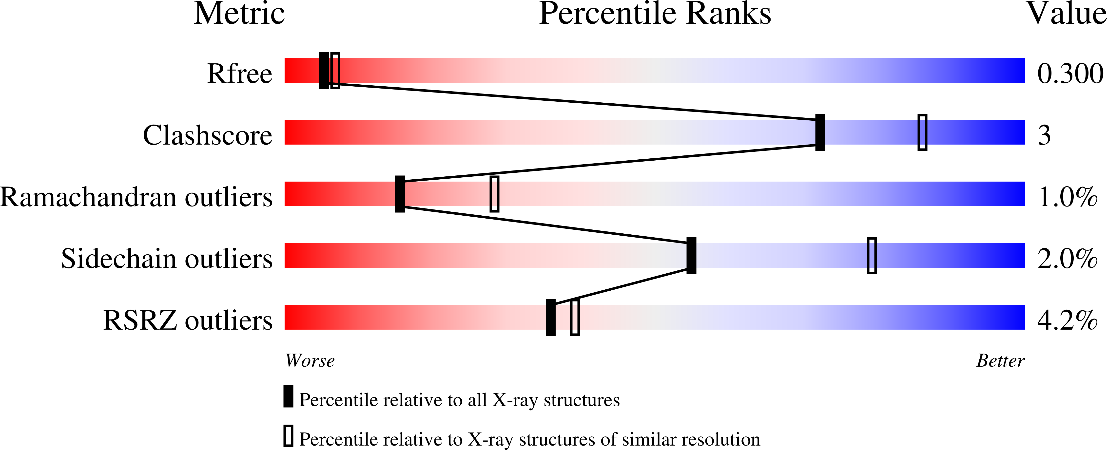
  • Method: X-RAY DIFFRACTION
  • Resolution: 2.50 Å
  • R-Value Free: 0.296 
  • R-Value Work: 0.238 
  • R-Value Observed: 0.241 

wwPDB Validation   3D Report Full Report


Ligand Structure Quality Assessment 


This is version 2.1 of the entry. See complete history


Literature

A designed supramolecular protein assembly with in vivo enzymatic activity.

Song, W.J.Tezcan, F.A.

(2014) Science 346: 1525-1528

  • DOI: https://doi.org/10.1126/science.1259680
  • Primary Citation of Related Structures:  
    4U9D, 4U9E

  • PubMed Abstract: 

    The generation of new enzymatic activities has mainly relied on repurposing the interiors of preexisting protein folds because of the challenge in designing functional, three-dimensional protein structures from first principles. Here we report an artificial metallo-β-lactamase, constructed via the self-assembly of a structurally and functionally unrelated, monomeric redox protein into a tetrameric assembly that possesses catalytic zinc sites in its interfaces. The designed metallo-β-lactamase is functional in the Escherichia coli periplasm and enables the bacteria to survive treatment with ampicillin. In vivo screening of libraries has yielded a variant that displays a catalytic proficiency [(k(cat)/K(m))/k(uncat)] for ampicillin hydrolysis of 2.3 × 10(6) and features the emergence of a highly mobile loop near the active site, a key component of natural β-lactamases to enable substrate interactions.


  • Organizational Affiliation

    Department of Chemistry and Biochemistry, University of California, San Diego, La Jolla, CA 92093-0356, USA.


Macromolecules
Find similar proteins by:  (by identity cutoff)  |  3D Structure
Entity ID: 1
MoleculeChains Sequence LengthOrganismDetailsImage
Soluble cytochrome b562
A, B, C, D
106Escherichia coliMutation(s): 14 
Gene Names: cybC
UniProt
Find proteins for P0ABE7 (Escherichia coli)
Explore P0ABE7 
Go to UniProtKB:  P0ABE7
Entity Groups  
Sequence Clusters30% Identity50% Identity70% Identity90% Identity95% Identity100% Identity
UniProt GroupP0ABE7
Sequence Annotations
Expand
  • Reference Sequence
Small Molecules
Ligands 2 Unique
IDChains Name / Formula / InChI Key2D Diagram3D Interactions
HEC
Query on HEC

Download Ideal Coordinates CCD File 
E [auth A],
K [auth B],
O [auth C],
P [auth D]
HEME C
C34 H34 Fe N4 O4
HXQIYSLZKNYNMH-LJNAALQVSA-N
ZN
Query on ZN

Download Ideal Coordinates CCD File 
F [auth A]
G [auth A]
H [auth A]
I [auth A]
J [auth A]
F [auth A],
G [auth A],
H [auth A],
I [auth A],
J [auth A],
L [auth B],
M [auth B],
N [auth B]
ZINC ION
Zn
PTFCDOFLOPIGGS-UHFFFAOYSA-N
Experimental Data & Validation

Experimental Data

  • Method: X-RAY DIFFRACTION
  • Resolution: 2.50 Å
  • R-Value Free: 0.296 
  • R-Value Work: 0.238 
  • R-Value Observed: 0.241 
  • Space Group: P 61
Unit Cell:
Length ( Å )Angle ( ˚ )
a = 54.83α = 90
b = 54.83β = 90
c = 237.82γ = 120
Software Package:
Software NamePurpose
REFMACrefinement
PDB_EXTRACTdata extraction

Structure Validation

View Full Validation Report


Ligand Structure Quality Assessment 


Entry History & Funding Information

Deposition Data


Funding OrganizationLocationGrant Number
National Science Foundation (NSF, United States)United StatesCHE1306646

Revision History  (Full details and data files)

  • Version 1.0: 2015-01-14
    Type: Initial release
  • Version 1.1: 2017-09-13
    Changes: Author supporting evidence, Database references, Derived calculations, Source and taxonomy, Structure summary
  • Version 1.2: 2019-11-27
    Changes: Author supporting evidence
  • Version 2.0: 2021-03-10
    Changes: Advisory, Atomic model, Data collection, Derived calculations, Non-polymer description, Structure summary
  • Version 2.1: 2023-12-27
    Changes: Data collection, Database references, Refinement description