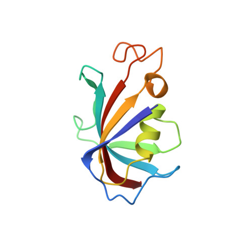
Structural basis of conformational transitions in the active site and 80's loop in the FK506-binding protein FKBP12.
Mustafi, S.M., Brecher, M., Zhang, J., Li, H., Lemaster, D.M., Hernandez, G.(2014) Biochem J 458: 525-536
- PubMed: 24405377
- DOI: https://doi.org/10.1042/BJ20131429
- Primary Citation of Related Structures:
4N19 - PubMed Abstract:
The extensive set of NMR doublings exhibited by the immunophilin FKBP12 (FK506-binding protein 12) arose from a slow transition to the cis-peptide configuration at Gly89 near the tip of the 80's loop, the site for numerous protein-recognition interactions for both FKBP12 and other FKBP domain proteins. The 80's loop also exhibited linebroadening, indicative of microsecond to millisecond conformational dynamics, but only in the trans-peptide state. The G89A variant shifted the trans-cis peptide equilibrium from 88:12 to 33:67, whereas a proline residue substitution induced fully the cis-peptide configuration. The 80's loop conformation in the G89P crystal structure at 1.50 Å resolution differed from wild-type FKBP12 primarily at residues 88, 89 and 90, and it closely resembled that reported for FKBP52. Structure-based chemical-shift predictions indicated that the microsecond to millisecond dynamics in the 80's loop probably arose from a concerted main chain (ψ88 and ϕ89) torsion angle transition. The indole side chain of Trp59 at the base of the active-site cleft was reoriented ~90o and the adjacent backbone was shifted in the G89P crystal structure. NOE analysis of wild-type FKBP12 demonstrated that this indole populates the perpendicular orientation at 20%. The 15N relaxation analysis was consistent with the indole reorientation occurring in the nanosecond timeframe. Recollection of the G89P crystal data at 1.20 Å resolution revealed a weaker wild-type-like orientation for the indole ring. Differences in the residues that underlie the Trp59 indole ring and altered interactions linking the 50's loop to the active site suggested that reorientation of this ring may be disfavoured in the other six members of the FKBP domain family that bear this active-site tryptophan residue.
Organizational Affiliation:
*Wadsworth Center, New York State Department of Health Empire State Plaza, Albany, NY 12201, U.S.A.