4K19

The structure of Human Siderocalin bound to the bacterial siderophore fluvibactin


Experimental Data Snapshot

  • Method: X-RAY DIFFRACTION
  • Resolution: 2.74 Å
  • R-Value Free: 0.294 
  • R-Value Work: 0.236 
  • R-Value Observed: 0.242 

wwPDB Validation   3D Report Full Report


Ligand Structure Quality Assessment 


This is version 1.2 of the entry. See complete history


Literature

Siderocalin Outwits the Coordination Chemistry of Vibriobactin, a Siderophore of Vibrio cholerae.

Allred, B.E.Correnti, C.Clifton, M.C.Strong, R.K.Raymond, K.N.

(2013) ACS Chem Biol 8: 1882-1887

  • DOI: https://doi.org/10.1021/cb4002552
  • Primary Citation of Related Structures:  
    4K19

  • PubMed Abstract: 

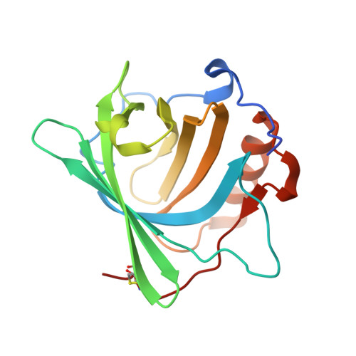
    The human protein siderocalin (Scn) inhibits bacterial iron acquisition by binding catechol siderophores. Several pathogenic bacteria respond by making stealth siderophores that are not recognized by Scn. Fluvibactin and vibriobactin, respectively of Vibrio fluvialis and Vibrio cholerae , include an oxazoline adjacent to a catechol. This chelating unit binds iron either in a catecholate or a phenolate-oxazoline coordination mode. The latter has been suggested to make vibriobactin a stealth siderophore without directly identifying the coordination mode in relation to Scn binding. We use Scn binding assays with the two siderophores and two oxazoline-substituted analogs and the crystal structure of Fe-fluvibactin:Scn to show that the oxazoline does not prevent Scn binding; hence, vibriobactin is not a stealth siderophore. We show that the phenolate-oxazoline coordination mode is present at physiological pH and is not bound by Scn. However, Scn binding shifts the coordination to the catecholate mode and thereby inactivates this siderophore.


  • Organizational Affiliation

    Department of Chemistry, University of California , Berkeley, California 94720-1460, United States.


Macromolecules
Find similar proteins by:  (by identity cutoff)  |  3D Structure
Entity ID: 1
MoleculeChains Sequence LengthOrganismDetailsImage
Neutrophil gelatinase-associated lipocalin
A, B, C
180Homo sapiensMutation(s): 1 
Gene Names: LCN2HNLNGAL
UniProt & NIH Common Fund Data Resources
Find proteins for P80188 (Homo sapiens)
Explore P80188 
Go to UniProtKB:  P80188
PHAROS:  P80188
GTEx:  ENSG00000148346 
Entity Groups  
Sequence Clusters30% Identity50% Identity70% Identity90% Identity95% Identity100% Identity
UniProt GroupP80188
Sequence Annotations
Expand
  • Reference Sequence
Small Molecules
Ligands 6 Unique
IDChains Name / Formula / InChI Key2D Diagram3D Interactions
1OD
Query on 1OD

Download Ideal Coordinates CCD File 
I [auth A],
K [auth B],
R [auth C]
(4S,5R)-N,N-bis{3-[(2,3-dihydroxybenzoyl)amino]propyl}-2-(2,3-dihydroxyphenyl)-5-methyl-4,5-dihydro-1,3-oxazole-4-carboxamide
C31 H34 N4 O10
LHJFXHHBUCCXIF-OSPHWJPCSA-N
SO4
Query on SO4

Download Ideal Coordinates CCD File 
F [auth A],
M [auth C]
SULFATE ION
O4 S
QAOWNCQODCNURD-UHFFFAOYSA-L
GOL
Query on GOL

Download Ideal Coordinates CCD File 
E [auth A],
H [auth A],
Q [auth C]
GLYCEROL
C3 H8 O3
PEDCQBHIVMGVHV-UHFFFAOYSA-N
FE
Query on FE

Download Ideal Coordinates CCD File 
D [auth A],
J [auth B],
L [auth C]
FE (III) ION
Fe
VTLYFUHAOXGGBS-UHFFFAOYSA-N
CL
Query on CL

Download Ideal Coordinates CCD File 
G [auth A],
N [auth C]
CHLORIDE ION
Cl
VEXZGXHMUGYJMC-UHFFFAOYSA-M
NA
Query on NA

Download Ideal Coordinates CCD File 
O [auth C],
P [auth C]
SODIUM ION
Na
FKNQFGJONOIPTF-UHFFFAOYSA-N
Binding Affinity Annotations 
IDSourceBinding Affinity
1OD PDBBind:  4K19 Kd: 4.4 (nM) from 1 assay(s)
BindingDB:  4K19 Kd: 34 (nM) from 1 assay(s)
Experimental Data & Validation

Experimental Data

  • Method: X-RAY DIFFRACTION
  • Resolution: 2.74 Å
  • R-Value Free: 0.294 
  • R-Value Work: 0.236 
  • R-Value Observed: 0.242 
  • Space Group: P 41 21 2
Unit Cell:
Length ( Å )Angle ( ˚ )
a = 114.2α = 90
b = 114.2β = 90
c = 119.3γ = 90
Software Package:
Software NamePurpose
DENZOdata reduction
SCALEPACKdata scaling
REFMACrefinement
PDB_EXTRACTdata extraction
HKL-2000data collection
HKL-2000data reduction
PHASERphasing

Structure Validation

View Full Validation Report


Ligand Structure Quality Assessment 


Entry History 

Deposition Data

Revision History  (Full details and data files)

  • Version 1.0: 2013-07-31
    Type: Initial release
  • Version 1.1: 2013-10-23
    Changes: Database references
  • Version 1.2: 2017-11-15
    Changes: Refinement description