4JKM

Crystal Structure of Clostridium perfringens beta-glucuronidase


Experimental Data Snapshot

  • Method: X-RAY DIFFRACTION
  • Resolution: 2.26 Å
  • R-Value Free: 0.234 
  • R-Value Work: 0.207 
  • R-Value Observed: 0.208 

wwPDB Validation   3D Report Full Report


This is version 1.2 of the entry. See complete history


Literature

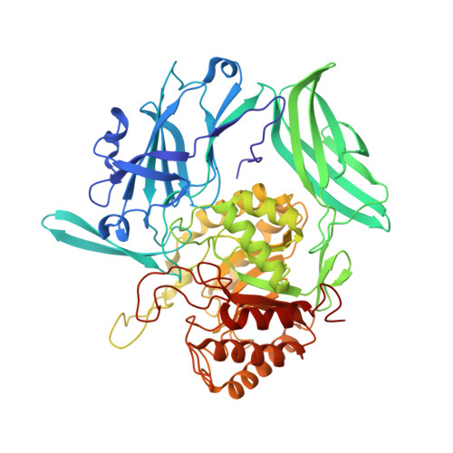
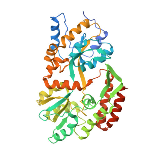
Structure and Inhibition of Microbiome beta-Glucuronidases Essential to the Alleviation of Cancer Drug Toxicity.

Wallace, B.D.Roberts, A.B.Pollet, R.M.Ingle, J.D.Biernat, K.A.Pellock, S.J.Venkatesh, M.K.Guthrie, L.O'Neal, S.K.Robinson, S.J.Dollinger, M.Figueroa, E.McShane, S.R.Cohen, R.D.Jin, J.Frye, S.V.Zamboni, W.C.Pepe-Ranney, C.Mani, S.Kelly, L.Redinbo, M.R.

(2015) Chem Biol 22: 1238-1249

  • DOI: https://doi.org/10.1016/j.chembiol.2015.08.005

  • PubMed Abstract: 

    The selective inhibition of bacterial β-glucuronidases was recently shown to alleviate drug-induced gastrointestinal toxicity in mice, including the damage caused by the widely used anticancer drug irinotecan. Here, we report crystal structures of representative β-glucuronidases from the Firmicutes Streptococcus agalactiae and Clostridium perfringens and the Proteobacterium Escherichia coli, and the characterization of a β-glucuronidase from the Bacteroidetes Bacteroides fragilis. While largely similar in structure, these enzymes exhibit marked differences in catalytic properties and propensities for inhibition, indicating that the microbiome maintains functional diversity in orthologous enzymes. Small changes in the structure of designed inhibitors can induce significant conformational changes in the β-glucuronidase active site. Finally, we establish that β-glucuronidase inhibition does not alter the serum pharmacokinetics of irinotecan or its metabolites in mice. Together, the data presented advance our in vitro and in vivo understanding of the microbial β-glucuronidases, a promising new set of targets for controlling drug-induced gastrointestinal toxicity.


  • Organizational Affiliation

    Department of Chemistry, University of North Carolina at Chapel Hill, NC 27599-3290, USA.


Macromolecules
Find similar proteins by:  (by identity cutoff)  |  3D Structure
Entity ID: 1
MoleculeChains Sequence LengthOrganismDetailsImage
Beta-glucuronidase
A, B
602Clostridium perfringens str. 13Mutation(s): 0 
Gene Names: bglR
EC: 3.2.1.31
UniProt
Find proteins for Q8XP19 (Clostridium perfringens (strain 13 / Type A))
Explore Q8XP19 
Go to UniProtKB:  Q8XP19
Entity Groups  
Sequence Clusters30% Identity50% Identity70% Identity90% Identity95% Identity100% Identity
UniProt GroupQ8XP19
Sequence Annotations
Expand
  • Reference Sequence
Find similar proteins by:  (by identity cutoff)  |  3D Structure
Entity ID: 2
MoleculeChains Sequence LengthOrganismDetailsImage
Maltose-binding periplasmic proteinC [auth D],
D [auth C]
400Escherichia coli K-12Mutation(s): 0 
Gene Names: b4034JW3994malE
UniProt
Find proteins for P0AEX9 (Escherichia coli (strain K12))
Explore P0AEX9 
Go to UniProtKB:  P0AEX9
Entity Groups  
Sequence Clusters30% Identity50% Identity70% Identity90% Identity95% Identity100% Identity
UniProt GroupP0AEX9
Sequence Annotations
Expand
  • Reference Sequence
Experimental Data & Validation

Experimental Data

  • Method: X-RAY DIFFRACTION
  • Resolution: 2.26 Å
  • R-Value Free: 0.234 
  • R-Value Work: 0.207 
  • R-Value Observed: 0.208 
  • Space Group: C 2 2 21
Unit Cell:
Length ( Å )Angle ( ˚ )
a = 71.511α = 90
b = 292.612β = 90
c = 239.907γ = 90
Software Package:
Software NamePurpose
XSCALEdata scaling
PHASERphasing
PHENIXrefinement
PDB_EXTRACTdata extraction
XDSdata scaling
XDSdata reduction

Structure Validation

View Full Validation Report


Entry History 

Deposition Data

Revision History  (Full details and data files)

  • Version 1.0: 2014-09-17
    Type: Initial release
  • Version 1.1: 2015-10-21
    Changes: Database references
  • Version 1.2: 2024-02-28
    Changes: Data collection, Database references