4FA4

Crystal Structure of WT MauG in Complex with Pre-Methylamine Dehydrogenase Aged 10 Days


Experimental Data Snapshot

  • Method: X-RAY DIFFRACTION
  • Resolution: 2.14 Å
  • R-Value Free: 0.227 
  • R-Value Work: 0.163 
  • R-Value Observed: 0.166 

Starting Model: experimental
View more details

wwPDB Validation   3D Report Full Report


Ligand Structure Quality Assessment 


This is version 1.5 of the entry. See complete history


Literature

Diradical intermediate within the context of tryptophan tryptophylquinone biosynthesis.

Yukl, E.T.Liu, F.Krzystek, J.Shin, S.Jensen, L.M.Davidson, V.L.Wilmot, C.M.Liu, A.

(2013) Proc Natl Acad Sci U S A 110: 4569-4573

  • DOI: https://doi.org/10.1073/pnas.1215011110
  • Primary Citation of Related Structures:  
    4FA1, 4FA4, 4FA5, 4FA9, 4FAN, 4FAV, 4FB1

  • PubMed Abstract: 

    Despite the importance of tryptophan (Trp) radicals in biology, very few radicals have been trapped and characterized in a physiologically meaningful context. Here we demonstrate that the diheme enzyme MauG uses Trp radical chemistry to catalyze formation of a Trp-derived tryptophan tryptophylquinone cofactor on its substrate protein, premethylamine dehydrogenase. The unusual six-electron oxidation that results in tryptophan tryptophylquinone formation occurs in three discrete two-electron catalytic steps. Here the exact order of these oxidation steps in the processive six-electron biosynthetic reaction is determined, and reaction intermediates are structurally characterized. The intermediates observed in crystal structures are also verified in solution using mass spectrometry. Furthermore, an unprecedented Trp-derived diradical species on premethylamine dehydrogenase, which is an intermediate in the first two-electron step, is characterized using high-frequency and -field electron paramagnetic resonance spectroscopy and UV-visible absorbance spectroscopy. This work defines a unique mechanism for radical-mediated catalysis of a protein substrate, and has broad implications in the areas of applied biocatalysis and understanding of oxidative protein modification during oxidative stress.


  • Organizational Affiliation

    Department of Biochemistry, Molecular Biology and Biophysics, University of Minnesota, Minneapolis, MN 55455, USA.


Macromolecules
Find similar proteins by:  (by identity cutoff)  |  3D Structure
Entity ID: 1
MoleculeChains Sequence LengthOrganismDetailsImage
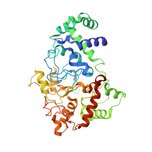
Methylamine utilization protein MauG
A, B
373Paracoccus denitrificans PD1222Mutation(s): 0 
Gene Names: mauGPden_4736
EC: 1
UniProt
Find proteins for Q51658 (Paracoccus denitrificans (strain Pd 1222))
Explore Q51658 
Go to UniProtKB:  Q51658
Entity Groups  
Sequence Clusters30% Identity50% Identity70% Identity90% Identity95% Identity100% Identity
UniProt GroupQ51658
Sequence Annotations
Expand
  • Reference Sequence
Find similar proteins by:  (by identity cutoff)  |  3D Structure
Entity ID: 2
MoleculeChains Sequence LengthOrganismDetailsImage
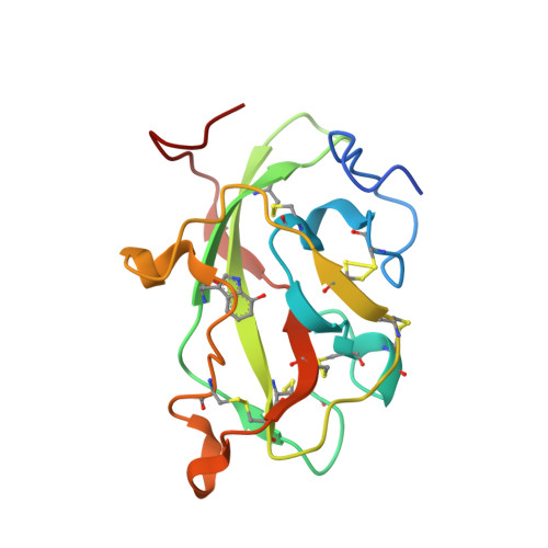
Methylamine dehydrogenase light chain
C, E
137Paracoccus denitrificansMutation(s): 0 
Gene Names: mauA
EC: 1.4.9.1
UniProt
Find proteins for P22619 (Paracoccus denitrificans)
Explore P22619 
Go to UniProtKB:  P22619
Entity Groups  
Sequence Clusters30% Identity50% Identity70% Identity90% Identity95% Identity100% Identity
UniProt GroupP22619
Sequence Annotations
Expand
  • Reference Sequence
Find similar proteins by:  (by identity cutoff)  |  3D Structure
Entity ID: 3
MoleculeChains Sequence LengthOrganismDetailsImage
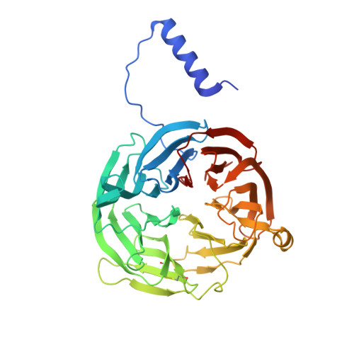
Methylamine dehydrogenase heavy chain
D, F
385Paracoccus denitrificans PD1222Mutation(s): 0 
Gene Names: Pden_4730
EC: 1.4.99.3 (PDB Primary Data), 1.4.9.1 (UniProt)
UniProt
Find proteins for A1BB97 (Paracoccus denitrificans (strain Pd 1222))
Explore A1BB97 
Go to UniProtKB:  A1BB97
Entity Groups  
Sequence Clusters30% Identity50% Identity70% Identity90% Identity95% Identity100% Identity
UniProt GroupA1BB97
Sequence Annotations
Expand
  • Reference Sequence
Small Molecules
Ligands 7 Unique
IDChains Name / Formula / InChI Key2D Diagram3D Interactions
HEC
Query on HEC

Download Ideal Coordinates CCD File 
H [auth A],
I [auth A],
N [auth B],
O [auth B]
HEME C
C34 H34 Fe N4 O4
HXQIYSLZKNYNMH-LJNAALQVSA-N
PGE
Query on PGE

Download Ideal Coordinates CCD File 
J [auth A]TRIETHYLENE GLYCOL
C6 H14 O4
ZIBGPFATKBEMQZ-UHFFFAOYSA-N
PO4
Query on PO4

Download Ideal Coordinates CCD File 
P [auth B]PHOSPHATE ION
O4 P
NBIIXXVUZAFLBC-UHFFFAOYSA-K
EDO
Query on EDO

Download Ideal Coordinates CCD File 
K [auth A],
Q [auth B]
1,2-ETHANEDIOL
C2 H6 O2
LYCAIKOWRPUZTN-UHFFFAOYSA-N
ACT
Query on ACT

Download Ideal Coordinates CCD File 
T [auth D],
U [auth F]
ACETATE ION
C2 H3 O2
QTBSBXVTEAMEQO-UHFFFAOYSA-M
CA
Query on CA

Download Ideal Coordinates CCD File 
G [auth A],
M [auth B]
CALCIUM ION
Ca
BHPQYMZQTOCNFJ-UHFFFAOYSA-N
NA
Query on NA

Download Ideal Coordinates CCD File 
L [auth A],
R [auth B],
S [auth B]
SODIUM ION
Na
FKNQFGJONOIPTF-UHFFFAOYSA-N
Modified Residues  1 Unique
IDChains TypeFormula2D DiagramParent
0AF
Query on 0AF
C, E
L-PEPTIDE LINKINGC11 H12 N2 O3TRP
Experimental Data & Validation

Experimental Data

  • Method: X-RAY DIFFRACTION
  • Resolution: 2.14 Å
  • R-Value Free: 0.227 
  • R-Value Work: 0.163 
  • R-Value Observed: 0.166 
  • Space Group: P 1
Unit Cell:
Length ( Å )Angle ( ˚ )
a = 55.53α = 109.92
b = 83.52β = 91.52
c = 107.78γ = 105.78
Software Package:
Software NamePurpose
Blu-Icedata collection
REFMACrefinement
HKL-2000data reduction
HKL-2000data scaling
REFMACphasing

Structure Validation

View Full Validation Report


Ligand Structure Quality Assessment 


Entry History 

Deposition Data

Revision History  (Full details and data files)

  • Version 1.0: 2013-03-06
    Type: Initial release
  • Version 1.1: 2013-03-27
    Changes: Database references
  • Version 1.2: 2013-04-17
    Changes: Database references
  • Version 1.3: 2023-09-13
    Changes: Data collection, Database references, Derived calculations, Refinement description
  • Version 1.4: 2023-12-06
    Changes: Data collection
  • Version 1.5: 2023-12-27
    Changes: Derived calculations