4B2T

The crystal structures of the eukaryotic chaperonin CCT reveal its functional partitioning


Experimental Data Snapshot

  • Method: X-RAY DIFFRACTION
  • Resolution: 5.50 Å
  • R-Value Free: 0.399 
  • R-Value Work: 0.340 
  • R-Value Observed: 0.340 

wwPDB Validation   3D Report Full Report


This is version 1.4 of the entry. See complete history

Re-refinement Note

This entry reflects an alternative modeling of the original data in: 2XSM


Literature

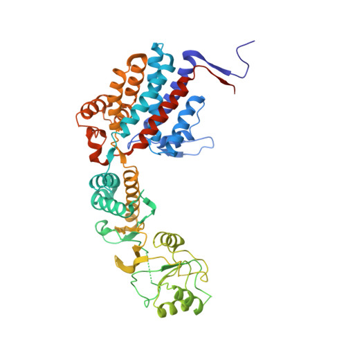
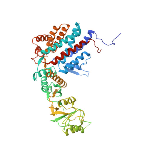
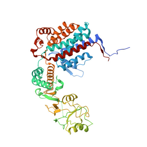
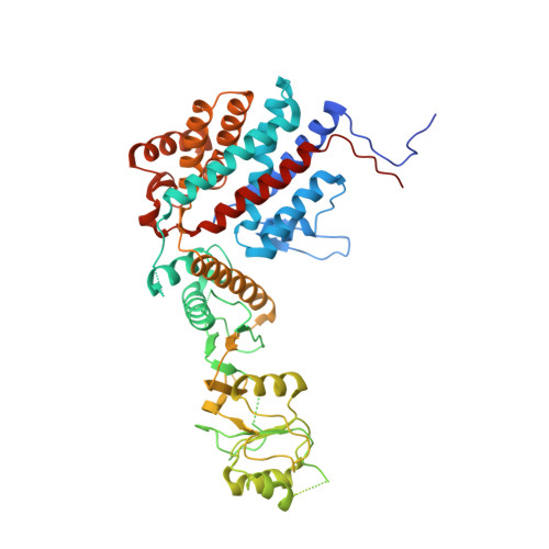
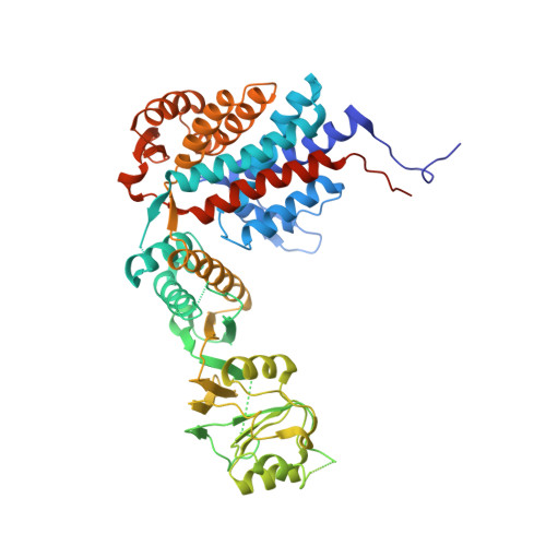
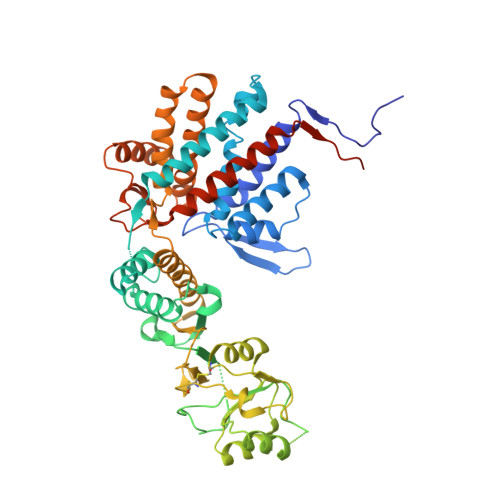
The Crystal Structures of the Eukaryotic Chaperonin Cct Reveal its Functional Partitioning

Kalisman, N.Schroder, G.F.Levitt, M.

(2013) Structure 21: 540

  • DOI: https://doi.org/10.1016/j.str.2013.01.017
  • Primary Citation of Related Structures:  
    4B2T, 4V8R

  • PubMed Abstract: 

    In eukaryotes, CCT is essential for the correct and efficient folding of many cytosolic proteins, most notably actin and tubulin. Structural studies of CCT have been hindered by the failure of standard crystallographic analysis to resolve its eight different subunit types at low resolutions. Here, we exhaustively assess the R value fit of all possible CCT models to available crystallographic data of the closed and open forms with resolutions of 3.8 Å and 5.5 Å, respectively. This unbiased analysis finds the native subunit arrangements with overwhelming significance. The resulting structures provide independent crystallographic proof of the subunit arrangement of CCT and map major asymmetrical features of the particle onto specific subunits. The actin and tubulin substrates both bind around subunit CCT6, which shows other structural anomalies. CCT is thus clearly partitioned, both functionally and evolutionary, into a substrate-binding side that is opposite to the ATP-hydrolyzing side.


  • Organizational Affiliation

    Department of Structural Biology, Stanford University School of Medicine, Stanford, CA 94305, USA.


Macromolecules
Find similar proteins by:  (by identity cutoff)  |  3D Structure
Entity ID: 1
MoleculeChains Sequence LengthOrganismDetailsImage
T-COMPLEX PROTEIN 1 SUBUNIT ALPHAA,
I [auth a]
556Bos taurusMutation(s): 0 
UniProt
Find proteins for Q32L40 (Bos taurus)
Explore Q32L40 
Go to UniProtKB:  Q32L40
Entity Groups  
Sequence Clusters30% Identity50% Identity70% Identity90% Identity95% Identity100% Identity
UniProt GroupQ32L40
Sequence Annotations
Expand
  • Reference Sequence
Find similar proteins by:  (by identity cutoff)  |  3D Structure
Entity ID: 2
MoleculeChains Sequence LengthOrganismDetailsImage
T-COMPLEX PROTEIN 1 SUBUNIT BETAB,
J [auth b]
535Bos taurusMutation(s): 0 
UniProt
Find proteins for Q3ZBH0 (Bos taurus)
Explore Q3ZBH0 
Go to UniProtKB:  Q3ZBH0
Entity Groups  
Sequence Clusters30% Identity50% Identity70% Identity90% Identity95% Identity100% Identity
UniProt GroupQ3ZBH0
Sequence Annotations
Expand
  • Reference Sequence
Find similar proteins by:  (by identity cutoff)  |  3D Structure
Entity ID: 3
MoleculeChains Sequence LengthOrganismDetailsImage
T-COMPLEX PROTEIN 1 SUBUNIT DELTAC [auth D],
K [auth d]
542Bos taurusMutation(s): 0 
UniProt
Find proteins for Q2T9X2 (Bos taurus)
Explore Q2T9X2 
Go to UniProtKB:  Q2T9X2
Entity Groups  
Sequence Clusters30% Identity50% Identity70% Identity90% Identity95% Identity100% Identity
UniProt GroupQ2T9X2
Sequence Annotations
Expand
  • Reference Sequence
Find similar proteins by:  (by identity cutoff)  |  3D Structure
Entity ID: 4
MoleculeChains Sequence LengthOrganismDetailsImage
T-COMPLEX PROTEIN 1 SUBUNIT EPSILOND [auth E],
L [auth e]
541Bos taurusMutation(s): 0 
UniProt
Find proteins for F1MWD3 (Bos taurus)
Explore F1MWD3 
Go to UniProtKB:  F1MWD3
Entity Groups  
Sequence Clusters30% Identity50% Identity70% Identity90% Identity95% Identity100% Identity
UniProt GroupF1MWD3
Sequence Annotations
Expand
  • Reference Sequence
Find similar proteins by:  (by identity cutoff)  |  3D Structure
Entity ID: 5
MoleculeChains Sequence LengthOrganismDetailsImage
T-COMPLEX PROTEIN 1 SUBUNIT GAMMAE [auth G],
M [auth g]
545Bos taurusMutation(s): 0 
UniProt
Find proteins for Q3T0K2 (Bos taurus)
Explore Q3T0K2 
Go to UniProtKB:  Q3T0K2
Entity Groups  
Sequence Clusters30% Identity50% Identity70% Identity90% Identity95% Identity100% Identity
UniProt GroupQ3T0K2
Sequence Annotations
Expand
  • Reference Sequence
Find similar proteins by:  (by identity cutoff)  |  3D Structure
Entity ID: 6
MoleculeChains Sequence LengthOrganismDetailsImage
T-COMPLEX PROTEIN 1 SUBUNIT ETAF [auth H],
N [auth h]
543Bos taurusMutation(s): 0 
UniProt
Find proteins for Q2NKZ1 (Bos taurus)
Explore Q2NKZ1 
Go to UniProtKB:  Q2NKZ1
Entity Groups  
Sequence Clusters30% Identity50% Identity70% Identity90% Identity95% Identity100% Identity
UniProt GroupQ2NKZ1
Sequence Annotations
Expand
  • Reference Sequence
Find similar proteins by:  (by identity cutoff)  |  3D Structure
Entity ID: 7
MoleculeChains Sequence LengthOrganismDetailsImage
T-COMPLEX PROTEIN 1 SUBUNIT THETAG [auth Q],
O [auth q]
548Bos taurusMutation(s): 0 
UniProt
Find proteins for Q3ZCI9 (Bos taurus)
Explore Q3ZCI9 
Go to UniProtKB:  Q3ZCI9
Entity Groups  
Sequence Clusters30% Identity50% Identity70% Identity90% Identity95% Identity100% Identity
UniProt GroupQ3ZCI9
Sequence Annotations
Expand
  • Reference Sequence
Find similar proteins by:  (by identity cutoff)  |  3D Structure
Entity ID: 8
MoleculeChains Sequence LengthOrganismDetailsImage
T-COMPLEX PROTEIN 1 SUBUNIT ZETAH [auth Z],
P [auth z]
531Bos taurusMutation(s): 0 
UniProt
Find proteins for Q3MHL7 (Bos taurus)
Explore Q3MHL7 
Go to UniProtKB:  Q3MHL7
Entity Groups  
Sequence Clusters30% Identity50% Identity70% Identity90% Identity95% Identity100% Identity
UniProt GroupQ3MHL7
Sequence Annotations
Expand
  • Reference Sequence
Experimental Data & Validation

Experimental Data

  • Method: X-RAY DIFFRACTION
  • Resolution: 5.50 Å
  • R-Value Free: 0.399 
  • R-Value Work: 0.340 
  • R-Value Observed: 0.340 
  • Space Group: P 21 21 2
Unit Cell:
Length ( Å )Angle ( ˚ )
a = 272.7α = 90
b = 313.5β = 90
c = 158.3γ = 90
Software Package:
Software NamePurpose
CNSrefinement

Structure Validation

View Full Validation Report


Entry History 

Deposition Data

Revision History  (Full details and data files)

  • Version 1.0: 2013-03-20
    Type: Initial release
  • Version 1.1: 2013-04-03
    Changes: Database references
  • Version 1.2: 2013-04-17
    Changes: Database references
  • Version 1.3: 2013-06-05
    Changes: Other, Structure summary
  • Version 1.4: 2019-11-06
    Changes: Data collection, Database references, Other