4A62

ParM from R1 plasmid in complex with peptide from C-terminus of ParR


Experimental Data Snapshot

  • Method: X-RAY DIFFRACTION
  • Resolution: 2.20 Å
  • R-Value Free: 0.269 
  • R-Value Work: 0.201 
  • R-Value Observed: 0.204 

wwPDB Validation   3D Report Full Report


Ligand Structure Quality Assessment 


This is version 1.3 of the entry. See complete history


Literature

A Bipolar Spindle of Antiparallel Parm Filaments Drives Bacterial Plasmid Segregation.

Gayathri, P.Fujii, T.Moller-Jensen, J.Van Den Ent, F.Namba, K.Lowe, J.

(2012) Science 338: 1334

  • DOI: https://doi.org/10.1126/science.1229091
  • Primary Citation of Related Structures:  
    4A61, 4A62, 4A6J

  • PubMed Abstract: 

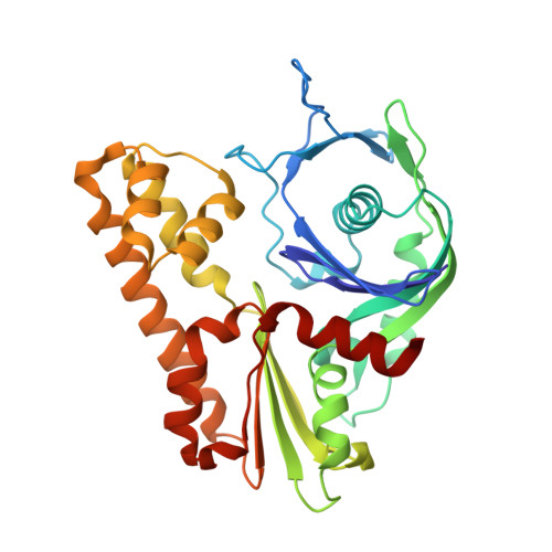
    To ensure their stable inheritance by daughter cells during cell division, bacterial low-copy-number plasmids make simple DNA segregating machines that use an elongating protein filament between sister plasmids. In the ParMRC system of the Escherichia coli R1 plasmid, ParM, an actinlike protein, forms the spindle between ParRC complexes on sister plasmids. By using a combination of structural work and total internal reflection fluorescence microscopy, we show that ParRC bound and could accelerate growth at only one end of polar ParM filaments, mechanistically resembling eukaryotic formins. The architecture of ParM filaments enabled two ParRC-bound filaments to associate in an antiparallel orientation, forming a bipolar spindle. The spindle elongated as a bundle of at least two antiparallel filaments, thereby pushing two plasmid clusters toward the poles.


  • Organizational Affiliation

    Medical Research Council Laboratory of Molecular Biology, Hills Road, Cambridge CB2 0QH, UK.


Macromolecules
Find similar proteins by:  (by identity cutoff)  |  3D Structure
Entity ID: 1
MoleculeChains Sequence LengthOrganismDetailsImage
PLASMID SEGREGATION PROTEIN PARM
A, B
320Escherichia coliMutation(s): 1 
UniProt
Find proteins for P11904 (Escherichia coli)
Explore P11904 
Go to UniProtKB:  P11904
Entity Groups  
Sequence Clusters30% Identity50% Identity70% Identity90% Identity95% Identity100% Identity
UniProt GroupP11904
Sequence Annotations
Expand
  • Reference Sequence

Find similar proteins by:  Sequence   |   3D Structure  

Entity ID: 2
MoleculeChains Sequence LengthOrganismDetailsImage
PROTEIN STBB
C, D
17Escherichia coliMutation(s): 0 
UniProt
Find proteins for P11906 (Escherichia coli)
Explore P11906 
Go to UniProtKB:  P11906
Entity Groups  
Sequence Clusters30% Identity50% Identity70% Identity90% Identity95% Identity100% Identity
UniProt GroupP11906
Sequence Annotations
Expand
  • Reference Sequence
Experimental Data & Validation

Experimental Data

  • Method: X-RAY DIFFRACTION
  • Resolution: 2.20 Å
  • R-Value Free: 0.269 
  • R-Value Work: 0.201 
  • R-Value Observed: 0.204 
  • Space Group: H 3 2
Unit Cell:
Length ( Å )Angle ( ˚ )
a = 146.85α = 90
b = 146.85β = 90
c = 171.32γ = 120
Software Package:
Software NamePurpose
PHENIXrefinement
MOSFLMdata reduction
SCALAdata scaling
PHASERphasing

Structure Validation

View Full Validation Report


Ligand Structure Quality Assessment 


Entry History 

Deposition Data

Revision History  (Full details and data files)

  • Version 1.0: 2012-11-07
    Type: Initial release
  • Version 1.1: 2012-12-19
    Changes: Database references
  • Version 1.2: 2018-01-24
    Changes: Source and taxonomy
  • Version 1.3: 2023-12-20
    Changes: Data collection, Database references, Derived calculations, Other, Refinement description