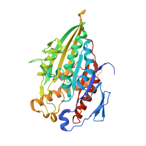
Structural Basis for the ATP-Induced Isomerization of Kinesin.
Chang, Q., Nitta, R., Inoue, S., Hirokawa, N.(2013) J Mol Biol 425: 1869
- PubMed: 23500491
- DOI: https://doi.org/10.1016/j.jmb.2013.03.004
- Primary Citation of Related Structures:
3ZFC, 3ZFD - PubMed Abstract:
Kinesin superfamily proteins (KIFs) are microtubule-based molecular motors driven by the energy derived from the hydrolysis of ATP. Previous studies have revealed that the ATP binding step is crucial both for the power stroke to produce motility and for the inter-domain regulation of ATPase activity to guarantee the processive movement of dimeric KIFs. Here, we report the first crystal structure of KIF4 complexed with the non-hydrolyzable ATP analog, AMPPNP (adenylyl imidodiphosphate), at 1.7Å resolution. By combining our structure with previously solved KIF1A structures complexed with two ATP analogs, molecular snapshots during ATP binding reveal that the closure of the nucleotide-binding pocket during ATP binding is achieved by closure of the backdoor. Closure of the backdoor stabilizes two mobile regions, switch I and switch II, to generate the phosphate tube from which hydrolyzed phosphate is released. Through the stabilization of switch II, the local conformational change at the catalytic center is further relayed to the neck-linker element that fully docks to the catalytic core to produce the power stroke. Because the neck linker is a sole element that connects the partner heads in dimeric KIFs, this tight structural coordination between the catalytic center and neck linker enables inter-domain communication between the partner heads. This study also revealed the putative microtubule-binding site of KIF4, thus providing structural insights that describe the specific binding of KIF4 to the microtubule.
Organizational Affiliation:
Department of Cell Biology and Anatomy, Graduate School of Medicine, The University of Tokyo, Hongo, Tokyo 113-0033, Japan; Department of Molecular Structure and Dynamics, Graduate School of Medicine, The University of Tokyo, Hongo, Tokyo 113-0033, Japan.


