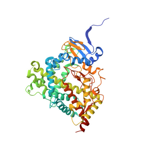
3RUK

Human Cytochrome P450 CYP17A1 in complex with Abiraterone


Experimental Data Snapshot

  • Method: X-RAY DIFFRACTION
  • Resolution: 2.60 Å
  • R-Value Free: 0.291 
  • R-Value Work: 0.218 
  • R-Value Observed: 0.222 

Starting Model: experimental
View more details

wwPDB Validation   3D Report Full Report


Ligand Structure Quality Assessment 


This is version 1.4 of the entry. See complete history


Literature

Structures of cytochrome P450 17A1 with prostate cancer drugs abiraterone and TOK-001.

Devore, N.M.Scott, E.E.

(2012) Nature 482: 116-119

  • DOI: https://doi.org/10.1038/nature10743
  • Primary Citation of Related Structures:  
    3RUK, 3SWZ

  • PubMed Abstract: 

    Cytochrome P450 17A1 (also known as CYP17A1 and cytochrome P450c17) catalyses the biosynthesis of androgens in humans. As prostate cancer cells proliferate in response to androgen steroids, CYP17A1 inhibition is a new strategy to prevent androgen synthesis and treat lethal metastatic castration-resistant prostate cancer, but drug development has been hampered by lack of information regarding the structure of CYP17A1. Here we report X-ray crystal structures of CYP17A1, which were obtained in the presence of either abiraterone, a first-in-class steroidal inhibitor recently approved by the US Food and Drug Administration for late-stage prostate cancer, or TOK-001, an inhibitor that is currently undergoing clinical trials. Both of these inhibitors bind the haem iron, forming a 60° angle above the haem plane and packing against the central I helix with the 3β-OH interacting with aspargine 202 in the F helix. Notably, this binding mode differs substantially from those that are predicted by homology models and from steroids in other cytochrome P450 enzymes with known structures, and some features of this binding mode are more similar to steroid receptors. Whereas the overall structure of CYP17A1 provides a rationale for understanding many mutations that are found in patients with steroidogenic diseases, the active site reveals multiple steric and hydrogen bonding features that will facilitate a better understanding of the enzyme's dual hydroxylase and lyase catalytic capabilities and assist in rational drug design. Specifically, structure-based design is expected to aid development of inhibitors that bind only CYP17A1 and solely inhibit its androgen-generating lyase activity to improve treatment of prostate and other hormone-responsive cancers.


  • Organizational Affiliation

    Department of Medicinal Chemistry, 1251 Wescoe Hall Drive, University of Kansas, Lawrence, Kansas 66045, USA.


Macromolecules
Find similar proteins by:  (by identity cutoff)  |  3D Structure
Entity ID: 1
MoleculeChains Sequence LengthOrganismDetailsImage
Steroid 17-alpha-hydroxylase/17,20 lyase
A, B, C, D
494Homo sapiensMutation(s): 0 
Gene Names: CYP17CYP17A1S17AH
EC: 1.14.99.9 (PDB Primary Data), 1.14.14.19 (UniProt), 1.14.14.32 (UniProt)
UniProt & NIH Common Fund Data Resources
Find proteins for P05093 (Homo sapiens)
Explore P05093 
Go to UniProtKB:  P05093
PHAROS:  P05093
GTEx:  ENSG00000148795 
Entity Groups  
Sequence Clusters30% Identity50% Identity70% Identity90% Identity95% Identity100% Identity
UniProt GroupP05093
Sequence Annotations
Expand
  • Reference Sequence
Small Molecules
Ligands 2 Unique
IDChains Name / Formula / InChI Key2D Diagram3D Interactions
HEM
Query on HEM

Download Ideal Coordinates CCD File 
E [auth A],
G [auth B],
I [auth C],
K [auth D]
PROTOPORPHYRIN IX CONTAINING FE
C34 H32 Fe N4 O4
KABFMIBPWCXCRK-RGGAHWMASA-L
AER
Query on AER

Download Ideal Coordinates CCD File 
F [auth A],
H [auth B],
J [auth C],
L [auth D]
Abiraterone
C24 H31 N O
GZOSMCIZMLWJML-VJLLXTKPSA-N
Binding Affinity Annotations 
IDSourceBinding Affinity
AER BindingDB:  3RUK IC50: min: 2, max: 800 (nM) from 11 assay(s)
Experimental Data & Validation

Experimental Data

  • Method: X-RAY DIFFRACTION
  • Resolution: 2.60 Å
  • R-Value Free: 0.291 
  • R-Value Work: 0.218 
  • R-Value Observed: 0.222 
  • Space Group: P 21 21 21
Unit Cell:
Length ( Å )Angle ( ˚ )
a = 85.813α = 90
b = 152.098β = 90
c = 172.882γ = 90
Software Package:
Software NamePurpose
Blu-Icedata collection
BALBESphasing
REFMACrefinement
MOSFLMdata reduction
SCALAdata scaling

Structure Validation

View Full Validation Report


Ligand Structure Quality Assessment 


Entry History 

Deposition Data

Revision History  (Full details and data files)

  • Version 1.0: 2012-01-25
    Type: Initial release
  • Version 1.1: 2012-02-01
    Changes: Database references
  • Version 1.2: 2017-11-08
    Changes: Refinement description
  • Version 1.3: 2024-02-28
    Changes: Data collection, Database references, Derived calculations
  • Version 1.4: 2024-04-03
    Changes: Refinement description