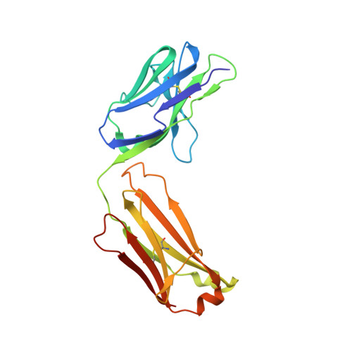
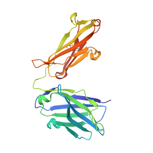
3QWO

Crystal structure of a motavizumab epitope-scaffold bound to motavizumab Fab


Experimental Data Snapshot

  • Method: X-RAY DIFFRACTION
  • Resolution: 1.90 Å
  • R-Value Free: 0.231 
  • R-Value Work: 0.188 
  • R-Value Observed: 0.190 

Starting Models: experimental
View more details

wwPDB Validation   3D Report Full Report


This is version 1.4 of the entry. See complete history


Literature

Design and characterization of epitope-scaffold immunogens that present the motavizumab epitope from respiratory syncytial virus.

McLellan, J.S.Correia, B.E.Chen, M.Yang, Y.Graham, B.S.Schief, W.R.Kwong, P.D.

(2011) J Mol Biol 409: 853-866

  • DOI: https://doi.org/10.1016/j.jmb.2011.04.044
  • Primary Citation of Related Structures:  
    3QWO

  • PubMed Abstract: 

    Respiratory syncytial virus (RSV) is a major cause of respiratory tract infections in infants, but an effective vaccine has not yet been developed. An ideal vaccine would elicit protective antibodies while avoiding virus-specific T-cell responses, which have been implicated in vaccine-enhanced disease with previous RSV vaccines. We propose that heterologous proteins designed to present RSV-neutralizing antibody epitopes and to elicit cognate antibodies have the potential to fulfill these vaccine requirements, as they can be fashioned to be free of viral T-cell epitopes. Here we present the design and characterization of three epitope-scaffolds that present the epitope of motavizumab, a potent neutralizing antibody that binds to a helix-loop-helix motif in the RSV fusion glycoprotein. Two of the epitope-scaffolds could be purified, and one epitope-scaffold based on a Staphylococcus aureus protein A domain bound motavizumab with kinetic and thermodynamic properties consistent with the free epitope-scaffold being stabilized in a conformation that closely resembled the motavizumab-bound state. This epitope-scaffold was well folded as assessed by circular dichroism and isothermal titration calorimetry, and its crystal structure (determined in complex with motavizumab to 1.9 Å resolution) was similar to the computationally designed model, with all hydrogen-bond interactions critical for binding to motavizumab preserved. Immunization of mice with this epitope-scaffold failed to elicit neutralizing antibodies but did elicit sera with F binding activity. The elicitation of F binding antibodies suggests that some of the design criteria for eliciting protective antibodies without virus-specific T-cell responses are being met, but additional optimization of these novel immunogens is required.


  • Organizational Affiliation

    Vaccine Research Center, National Institute of Allergy and Infectious Diseases, National Institutes of Health, Bethesda, MD 20892, USA. mclellanja@mail.nih.gov


Macromolecules
Find similar proteins by:  (by identity cutoff)  |  3D Structure
Entity ID: 1
MoleculeChains Sequence LengthOrganismDetailsImage
motavizumab light chainA [auth L],
C [auth B]
213Mus musculusMutation(s): 0 
Entity Groups  
Sequence Clusters30% Identity50% Identity70% Identity90% Identity95% Identity100% Identity
Sequence Annotations
Expand
  • Reference Sequence
Find similar proteins by:  (by identity cutoff)  |  3D Structure
Entity ID: 2
MoleculeChains Sequence LengthOrganismDetailsImage
motavizumab heavy chainB [auth H],
D [auth A]
225Mus musculusMutation(s): 0 
Entity Groups  
Sequence Clusters30% Identity50% Identity70% Identity90% Identity95% Identity100% Identity
Sequence Annotations
Expand
  • Reference Sequence
Find similar proteins by:  (by identity cutoff)  |  3D Structure
Entity ID: 3
MoleculeChains Sequence LengthOrganismDetailsImage
motavizumab epitope scaffoldE [auth C],
F [auth P]
57Staphylococcus aureusMutation(s): 0 
Entity Groups  
Sequence Clusters30% Identity50% Identity70% Identity90% Identity95% Identity100% Identity
Sequence Annotations
Expand
  • Reference Sequence
Experimental Data & Validation

Experimental Data

  • Method: X-RAY DIFFRACTION
  • Resolution: 1.90 Å
  • R-Value Free: 0.231 
  • R-Value Work: 0.188 
  • R-Value Observed: 0.190 
  • Space Group: P 1 21 1
Unit Cell:
Length ( Å )Angle ( ˚ )
a = 75.762α = 90
b = 74.425β = 101.75
c = 86.398γ = 90
Software Package:
Software NamePurpose
SERGUIdata collection
PHASERphasing
PHENIXrefinement
HKL-2000data reduction
HKL-2000data scaling

Structure Validation

View Full Validation Report


Entry History 

Deposition Data

Revision History  (Full details and data files)

  • Version 1.0: 2011-05-04
    Type: Initial release
  • Version 1.1: 2011-07-13
    Changes: Version format compliance
  • Version 1.2: 2011-08-10
    Changes: Database references
  • Version 1.3: 2023-09-13
    Changes: Data collection, Database references, Derived calculations, Refinement description
  • Version 1.4: 2024-11-06
    Changes: Structure summary