


Crystal structure of PNP with an inhibitor DADME_immH from Vibrio cholerae
Kim, J., Ramagopal, U.A., Burley, S.K., Almo, S.C.To be published.
Experimental Data Snapshot
Starting Model: experimental
View more details
Entity ID: 1 | |||||
|---|---|---|---|---|---|
| Molecule | Chains | Sequence Length | Organism | Details | Image |
| Purine nucleoside phosphorylase deoD-type 1 | 241 | Vibrio cholerae | Mutation(s): 0 Gene Names: deoD, deoD1, VC_2347 EC: 2.4.2.1 | 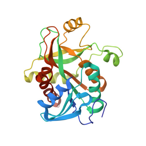 | |
UniProt | |||||
Find proteins for Q9KPM0 (Vibrio cholerae serotype O1 (strain ATCC 39315 / El Tor Inaba N16961)) Explore Q9KPM0 Go to UniProtKB: Q9KPM0 | |||||
Entity Groups | |||||
| Sequence Clusters | 30% Identity50% Identity70% Identity90% Identity95% Identity100% Identity | ||||
| UniProt Group | Q9KPM0 | ||||
Sequence AnnotationsExpand | |||||
| |||||
| Ligands 2 Unique | |||||
|---|---|---|---|---|---|
| ID | Chains | Name / Formula / InChI Key | 2D Diagram | 3D Interactions | |
| DIH Query on DIH | AA [auth H] CA [auth I] EA [auth J] GA [auth K] IA [auth L] | 7-[[(3R,4R)-3-(hydroxymethyl)-4-oxidanyl-pyrrolidin-1-ium-1-yl]methyl]-3,5-dihydropyrrolo[3,2-d]pyrimidin-4-one C12 H17 N4 O3 AFNHHLILYQEHKK-BDAKNGLRSA-O |  | ||
| PO4 Query on PO4 | BA [auth H] DA [auth I] FA [auth J] HA [auth K] JA [auth L] | PHOSPHATE ION O4 P NBIIXXVUZAFLBC-UHFFFAOYSA-K |  | ||
| Length ( Å ) | Angle ( ˚ ) |
|---|---|
| a = 101.375 | α = 90 |
| b = 160.974 | β = 90 |
| c = 188.685 | γ = 90 |
| Software Name | Purpose |
|---|---|
| HKL-2000 | data collection |
| MOLREP | phasing |
| REFMAC | refinement |
| HKL-2000 | data reduction |
| HKL-2000 | data scaling |
RCSB PDB (citation) is hosted by
RCSB PDB is a member of the

