3N7R

Crystal structure of the ectodomain complex of the CGRP receptor, a Class-B GPCR, reveals the site of drug antagonism


Experimental Data Snapshot

  • Method: X-RAY DIFFRACTION
  • Resolution: 2.90 Å
  • R-Value Free: 0.267 
  • R-Value Work: 0.232 
  • R-Value Observed: 0.234 

wwPDB Validation   3D Report Full Report


This is version 1.3 of the entry. See complete history


Literature

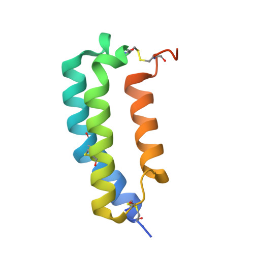
Crystal Structure of the Ectodomain Complex of the CGRP Receptor, a Class-B GPCR, Reveals the Site of Drug Antagonism.

Ter Haar, E.Koth, C.M.Abdul-Manan, N.Swenson, L.Coll, J.T.Lippke, J.A.Lepre, C.A.Garcia-Guzman, M.Moore, J.M.

(2010) Structure 18: 1083-1093

  • DOI: https://doi.org/10.1016/j.str.2010.05.014
  • Primary Citation of Related Structures:  
    3N7P, 3N7R, 3N7S

  • PubMed Abstract: 

    Dysregulation of the calcitonin gene-related peptide (CGRP), a potent vasodilator, is directly implicated in the pathogenesis of migraine. CGRP binds to and signals through the CGRP receptor (CGRP-R), a heterodimer containing the calcitonin receptor-like receptor (CLR), a class B GPCR, and RAMP1, a receptor activity-modifying protein. We have solved the crystal structure of the CLR/RAMP1 N-terminal ectodomain heterodimer, revealing how RAMPs bind to and potentially modulate the activities of the CLR GPCR subfamily. We also report the structures of CLR/RAMP1 in complex with the clinical receptor antagonists olcegepant (BIBN4096BS) and telcagepant (MK0974). Both drugs act by blocking access to the peptide-binding cleft at the interface of CLR and RAMP1. These structures illustrate, for the first time, how small molecules bind to and modulate the activity of a class B GPCR, and highlight the challenges of designing potent receptor antagonists for the treatment of migraine and other class B GPCR-related diseases.


  • Organizational Affiliation

    Vertex Pharmaceuticals Incorporated, 130 Waverly Street, Cambridge, MA 02139, USA.


Macromolecules
Find similar proteins by:  (by identity cutoff)  |  3D Structure
Entity ID: 1
MoleculeChains Sequence LengthOrganismDetailsImage
Calcitonin gene-related peptide type 1 receptor
A, B
115Homo sapiensMutation(s): 0 
Gene Names: CALCRLCALRL_HUMANCGRPR
UniProt & NIH Common Fund Data Resources
Find proteins for Q16602 (Homo sapiens)
Explore Q16602 
Go to UniProtKB:  Q16602
PHAROS:  Q16602
GTEx:  ENSG00000064989 
Entity Groups  
Sequence Clusters30% Identity50% Identity70% Identity90% Identity95% Identity100% Identity
UniProt GroupQ16602
Sequence Annotations
Expand
  • Reference Sequence
Find similar proteins by:  (by identity cutoff)  |  3D Structure
Entity ID: 2
MoleculeChains Sequence LengthOrganismDetailsImage
Receptor activity-modifying protein 1
C, D
96Homo sapiensMutation(s): 0 
Gene Names: RAMP1RAMP1_HUMAN
UniProt & NIH Common Fund Data Resources
Find proteins for O60894 (Homo sapiens)
Explore O60894 
Go to UniProtKB:  O60894
PHAROS:  O60894
GTEx:  ENSG00000132329 
Entity Groups  
Sequence Clusters30% Identity50% Identity70% Identity90% Identity95% Identity100% Identity
UniProt GroupO60894
Sequence Annotations
Expand
  • Reference Sequence
Small Molecules
Ligands 2 Unique
IDChains Name / Formula / InChI Key2D Diagram3D Interactions
3N6
Query on 3N6

Download Ideal Coordinates CCD File 
E [auth A]N-{(1S)-5-amino-1-[(4-pyridin-4-ylpiperazin-1-yl)carbonyl]pentyl}-3,5-dibromo-Nalpha-{[4-(2-oxo-1,4-dihydroquinazolin-3 (2H)-yl)piperidin-1-yl]carbonyl}-D-tyrosinamide
C38 H47 Br2 N9 O5
ITIXDWVDFFXNEG-JHOUSYSJSA-N
N7R
Query on N7R

Download Ideal Coordinates CCD File 
F [auth B]N-[(3R,6S)-6-(2,3-difluorophenyl)-2-oxo-1-(2,2,2-trifluoroethyl)azepan-3-yl]-4-(2-oxo-2,3-dihydro-1H-imidazo[4,5-b]pyridin-1-yl)piperidine-1-carboxamide
C26 H27 F5 N6 O3
CGDZXLJGHVKVIE-DNVCBOLYSA-N
Binding Affinity Annotations 
IDSourceBinding Affinity
N7R BindingDB:  3N7R Ki: min: 0.77, max: 0.8 (nM) from 2 assay(s)
Kd: 1.9 (nM) from 1 assay(s)
IC50: min: 2, max: 11 (nM) from 4 assay(s)
PDBBind:  3N7R Kd: 20 (nM) from 1 assay(s)
3N6 BindingDB:  3N7R Ki: min: 5.00e-3, max: 3.4 (nM) from 5 assay(s)
IC50: min: 0.02, max: 4 (nM) from 4 assay(s)
EC50: 0.03 (nM) from 1 assay(s)
Experimental Data & Validation

Experimental Data

  • Method: X-RAY DIFFRACTION
  • Resolution: 2.90 Å
  • R-Value Free: 0.267 
  • R-Value Work: 0.232 
  • R-Value Observed: 0.234 
  • Space Group: C 2 2 21
Unit Cell:
Length ( Å )Angle ( ˚ )
a = 77.94α = 90
b = 118.2β = 90
c = 133.26γ = 90
Software Package:
Software NamePurpose
DENZOdata reduction
SCALEPACKdata scaling
BUSTER-TNTrefinement
PDB_EXTRACTdata extraction
HKL-2000data collection
HKL-2000data reduction
BUSTERrefinement

Structure Validation

View Full Validation Report


Entry History 

Deposition Data

  • Released Date: 2010-09-15 
  • Deposition Author(s): Ter Haar, E.

Revision History  (Full details and data files)

  • Version 1.0: 2010-09-15
    Type: Initial release
  • Version 1.1: 2011-07-13
    Changes: Version format compliance
  • Version 1.2: 2017-11-08
    Changes: Refinement description
  • Version 1.3: 2022-10-26
    Changes: Database references, Derived calculations, Structure summary