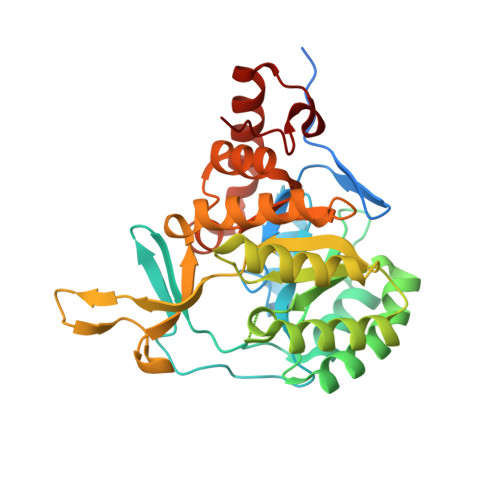
3MJY

Crystal structure of dihydroorotate dehydrogenase from Leishmania major in complex with 5-Aminoorotic acid


Experimental Data Snapshot

  • Method: X-RAY DIFFRACTION
  • Resolution: 1.96 Å
  • R-Value Free: 0.198 
  • R-Value Work: 0.158 
  • R-Value Observed: 0.160 

Starting Model: experimental
View more details

wwPDB Validation   3D Report Full Report


Ligand Structure Quality Assessment 


This is version 1.2 of the entry. See complete history


Literature

Novel insights for dihydroorotate dehydrogenase class 1A inhibitors discovery.

Cheleski, J.Rocha, J.R.Pinheiro, M.P.Wiggers, H.J.da Silva, A.B.Nonato, M.C.Montanari, C.A.

(2010) Eur J Med Chem 45: 5899-5909

  • DOI: https://doi.org/10.1016/j.ejmech.2010.09.055
  • Primary Citation of Related Structures:  
    3MHU, 3MJY

  • PubMed Abstract: 

    The enzyme dihydroorotate dehydrogenase (DHODH) has been suggested as a promising target for the design of trypanocidal agents. We report here the discovery of novel inhibitors of Trypanosoma cruzi DHODH identified by a combination of virtual screening and ITC methods. Monitoring of the enzymatic reaction in the presence of selected ligands together with structural information obtained from X-ray crystallography analysis have allowed the identification and validation of a novel site of interaction (S2 site). This has provided important structural insights for the rational design of T. cruzi and Leishmania major DHODH inhibitors. The most potent compound (1) in the investigated series inhibits TcDHODH enzyme with Kiapp value of 19.28 μM and possesses a ligand efficiency of 0.54 kcal mol(-1) per non-H atom. The compounds described in this work are promising hits for further development.


  • Organizational Affiliation

    Grupo de Estudos em Química Medicinal de Produtos Naturais, NEQUIMED-PN, Instituto de Química de São Carlos, Universidade de São Paulo, Av. Trabalhador Sancarlense 400, 13560-970, São Carlos-SP, Brazil.


Macromolecules
Find similar proteins by:  (by identity cutoff)  |  3D Structure
Entity ID: 1
MoleculeChains Sequence LengthOrganismDetailsImage
Dihydroorotate dehydrogenase
A, B
346Leishmania majorMutation(s): 0 
Gene Names: LmjF16.0530
EC: 1.3.3.1 (PDB Primary Data), 1.3.98.1 (UniProt)
UniProt
Find proteins for Q4QEW7 (Leishmania major)
Explore Q4QEW7 
Go to UniProtKB:  Q4QEW7
Entity Groups  
Sequence Clusters30% Identity50% Identity70% Identity90% Identity95% Identity100% Identity
UniProt GroupQ4QEW7
Sequence Annotations
Expand
  • Reference Sequence
Small Molecules
Ligands 4 Unique
IDChains Name / Formula / InChI Key2D Diagram3D Interactions
FMN
Query on FMN

Download Ideal Coordinates CCD File 
C [auth A],
I [auth B]
FLAVIN MONONUCLEOTIDE
C17 H21 N4 O9 P
FVTCRASFADXXNN-SCRDCRAPSA-N
IJZ
Query on IJZ

Download Ideal Coordinates CCD File 
D [auth A],
J [auth B]
5-amino-2,6-dioxo-1,2,3,6-tetrahydropyrimidine-4-carboxylic acid
C5 H5 N3 O4
HWCXJKLFOSBVLH-UHFFFAOYSA-N
SO4
Query on SO4

Download Ideal Coordinates CCD File 
H [auth A]SULFATE ION
O4 S
QAOWNCQODCNURD-UHFFFAOYSA-L
GOL
Query on GOL

Download Ideal Coordinates CCD File 
E [auth A],
F [auth A],
G [auth A],
K [auth B],
L [auth B]
GLYCEROL
C3 H8 O3
PEDCQBHIVMGVHV-UHFFFAOYSA-N
Experimental Data & Validation

Experimental Data

  • Method: X-RAY DIFFRACTION
  • Resolution: 1.96 Å
  • R-Value Free: 0.198 
  • R-Value Work: 0.158 
  • R-Value Observed: 0.160 
  • Space Group: P 61
Unit Cell:
Length ( Å )Angle ( ˚ )
a = 141.264α = 90
b = 141.264β = 90
c = 68.743γ = 120
Software Package:
Software NamePurpose
MAR345dtbdata collection
MOLREPphasing
REFMACrefinement
MOSFLMdata reduction
SCALAdata scaling

Structure Validation

View Full Validation Report


Ligand Structure Quality Assessment 


Entry History 

Deposition Data

Revision History  (Full details and data files)

  • Version 1.0: 2011-02-23
    Type: Initial release
  • Version 1.1: 2011-07-13
    Changes: Version format compliance
  • Version 1.2: 2023-09-06
    Changes: Data collection, Database references, Derived calculations, Refinement description