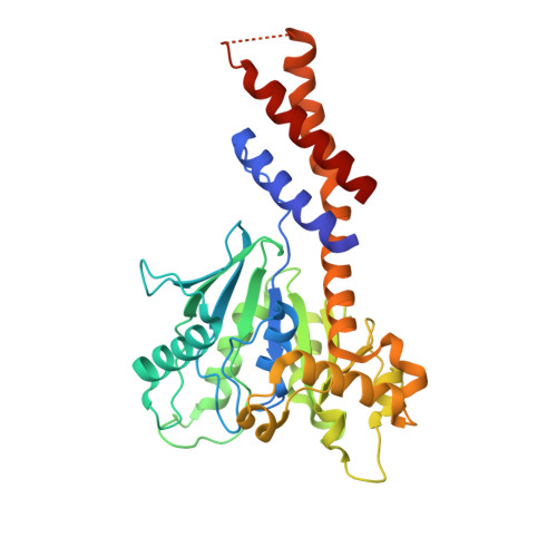
3ZYC

DYNAMIN 1 GTPASE GED FUSION DIMER COMPLEXED WITH GMPPCP


Experimental Data Snapshot

  • Method: X-RAY DIFFRACTION
  • Resolution: 2.20 Å
  • R-Value Free: 0.269 
  • R-Value Work: 0.225 
  • R-Value Observed: 0.225 

Starting Model: experimental
View more details

wwPDB Validation   3D Report Full Report


Ligand Structure Quality Assessment 


This is version 1.4 of the entry. See complete history


Literature

A Pseudoatomic Model of the Dynamin Polymer Identifies a Hydrolysis-Dependent Powerstroke.

Chappie, J.S.Mears, J.A.Fang, S.Leonard, M.Schmid, S.L.Milligan, R.A.Hinshaw, J.E.Dyda, F.

(2011) Cell 147: 209

  • DOI: https://doi.org/10.1016/j.cell.2011.09.003
  • Primary Citation of Related Structures:  
    3ZYC, 3ZYS

  • PubMed Abstract: 

    The GTPase dynamin catalyzes membrane fission by forming a collar around the necks of clathrin-coated pits, but the specific structural interactions and conformational changes that drive this process remain a mystery. We present the GMPPCP-bound structures of the truncated human dynamin 1 helical polymer at 12.2 Å and a fusion protein, GG, linking human dynamin 1's catalytic G domain to its GTPase effector domain (GED) at 2.2 Å. The structures reveal the position and connectivity of dynamin fragments in the assembled structure, showing that G domain dimers only form between tetramers in sequential rungs of the dynamin helix. Using chemical crosslinking, we demonstrate that dynamin tetramers are made of two dimers, in which the G domain of one molecule interacts in trans with the GED of another. Structural comparison of GG(GMPPCP) to the GG transition-state complex identifies a hydrolysis-dependent powerstroke that may play a role in membrane-remodeling events necessary for fission.


  • Organizational Affiliation

    Laboratory of Molecular Biology, National Institute of Diabetes and Digestive and Kidney Diseases, NIH, Bethesda, MD 20892, USA.


Macromolecules
Find similar proteins by:  (by identity cutoff)  |  3D Structure
Entity ID: 1
MoleculeChains Sequence LengthOrganismDetailsImage
DYNAMIN-1A,
B [auth D]
353Homo sapiensMutation(s): 1 
EC: 3.6.5.5
UniProt & NIH Common Fund Data Resources
Find proteins for Q05193 (Homo sapiens)
Explore Q05193 
Go to UniProtKB:  Q05193
PHAROS:  Q05193
GTEx:  ENSG00000106976 
Entity Groups  
Sequence Clusters30% Identity50% Identity70% Identity90% Identity95% Identity100% Identity
UniProt GroupQ05193
Sequence Annotations
Expand
  • Reference Sequence
Experimental Data & Validation

Experimental Data

  • Method: X-RAY DIFFRACTION
  • Resolution: 2.20 Å
  • R-Value Free: 0.269 
  • R-Value Work: 0.225 
  • R-Value Observed: 0.225 
  • Space Group: P 1 21 1
Unit Cell:
Length ( Å )Angle ( ˚ )
a = 44.151α = 90
b = 82.635β = 96.68
c = 95.014γ = 90
Software Package:
Software NamePurpose
CNSrefinement
XDSdata reduction
XDSdata scaling
PHASERphasing

Structure Validation

View Full Validation Report


Ligand Structure Quality Assessment 


Entry History 

Deposition Data

Revision History  (Full details and data files)

  • Version 1.0: 2011-10-12
    Type: Initial release
  • Version 1.1: 2017-06-28
    Changes: Data collection
  • Version 1.2: 2019-05-08
    Changes: Data collection, Experimental preparation
  • Version 1.3: 2019-05-15
    Changes: Data collection, Experimental preparation
  • Version 1.4: 2023-12-20
    Changes: Data collection, Database references, Derived calculations, Other, Refinement description