3ZKJ

Crystal Structure of Ankyrin Repeat and Socs Box-Containing Protein 9 (Asb9) in Complex with Elonginb and Elonginc


Experimental Data Snapshot

  • Method: X-RAY DIFFRACTION
  • Resolution: 2.58 Å
  • R-Value Free: 0.265 
  • R-Value Work: 0.227 
  • R-Value Observed: 0.229 

wwPDB Validation   3D Report Full Report


This is version 1.4 of the entry. See complete history


Literature

Molecular Architecture of the Ankyrin Socs Box Family of Cul5-Dependent E3 Ubiquitin Ligases

Muniz, J.R.C.Guo, K.Kershaw, N.J.Ayinampudi, V.von Delft, F.Babon, J.J.Bullock, A.N.

(2013) J Mol Biol 425: 3166

  • DOI: https://doi.org/10.1016/j.jmb.2013.06.015
  • Primary Citation of Related Structures:  
    2WZK, 3ZKJ

  • PubMed Abstract: 

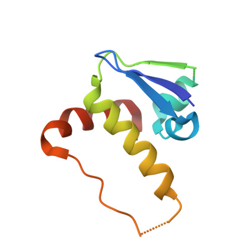
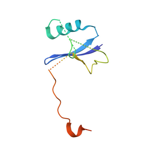
    Multi-subunit Cullin-RING E3 ligases often use repeat domain proteins as substrate-specific adaptors. Structures of these macromolecular assemblies are determined for the F-box-containing leucine-rich repeat and WD40 repeat families, but not for the suppressor of cytokine signaling (SOCS)-box-containing ankyrin repeat proteins (ASB1-18), which assemble with Elongins B and C and Cul5. We determined the crystal structures of the ternary complex of ASB9-Elongin B/C as well as the interacting N-terminal domain of Cul5 and used structural comparisons to establish a model for the complete Cul5-based E3 ligase. The structures reveal a distinct architecture of the ASB9 complex that positions the ankyrin domain coaxial to the SOCS box-Elongin B/C complex and perpendicular to other repeat protein complexes. This alternative architecture appears favorable to present the ankyrin domain substrate-binding site to the E2-ubiquitin, while also providing spacing suitable for bulky ASB9 substrates, such as the creatine kinases. The presented Cul5 structure also differs from previous models and deviates from other Cullins via a rigid-body rotation between Cullin repeats. This work highlights the adaptability of repeat domain proteins as scaffolds in substrate recognition and lays the foundation for future structure-function studies of this important E3 family.


  • Organizational Affiliation

    Structural Genomics Consortium, University of Oxford, Old Road Campus Research Building, Roosevelt Drive, Oxford OX3 7DQ, United Kingdom.


Macromolecules
Find similar proteins by:  (by identity cutoff)  |  3D Structure
Entity ID: 1
MoleculeChains Sequence LengthOrganismDetailsImage
ANKYRIN REPEAT AND SOCS BOX PROTEIN 9
A, D
261Homo sapiensMutation(s): 0 
UniProt & NIH Common Fund Data Resources
Find proteins for Q96DX5 (Homo sapiens)
Explore Q96DX5 
Go to UniProtKB:  Q96DX5
PHAROS:  Q96DX5
GTEx:  ENSG00000102048 
Entity Groups  
Sequence Clusters30% Identity50% Identity70% Identity90% Identity95% Identity100% Identity
UniProt GroupQ96DX5
Sequence Annotations
Expand
  • Reference Sequence
Find similar proteins by:  (by identity cutoff)  |  3D Structure
Entity ID: 2
MoleculeChains Sequence LengthOrganismDetailsImage
TRANSCRIPTION ELONGATION FACTOR B POLYPEPTIDE 1
B, E
96Homo sapiensMutation(s): 0 
UniProt & NIH Common Fund Data Resources
Find proteins for Q15369 (Homo sapiens)
Explore Q15369 
Go to UniProtKB:  Q15369
PHAROS:  Q15369
GTEx:  ENSG00000154582 
Entity Groups  
Sequence Clusters30% Identity50% Identity70% Identity90% Identity95% Identity100% Identity
UniProt GroupQ15369
Sequence Annotations
Expand
  • Reference Sequence
Find similar proteins by:  (by identity cutoff)  |  3D Structure
Entity ID: 3
MoleculeChains Sequence LengthOrganismDetailsImage
TRANSCRIPTION ELONGATION FACTOR B POLYPEPTIDE 2
C, F
118Homo sapiensMutation(s): 0 
UniProt & NIH Common Fund Data Resources
Find proteins for Q15370 (Homo sapiens)
Explore Q15370 
Go to UniProtKB:  Q15370
PHAROS:  Q15370
GTEx:  ENSG00000103363 
Entity Groups  
Sequence Clusters30% Identity50% Identity70% Identity90% Identity95% Identity100% Identity
UniProt GroupQ15370
Sequence Annotations
Expand
  • Reference Sequence
Experimental Data & Validation

Experimental Data

  • Method: X-RAY DIFFRACTION
  • Resolution: 2.58 Å
  • R-Value Free: 0.265 
  • R-Value Work: 0.227 
  • R-Value Observed: 0.229 
  • Space Group: P 21 2 21
Unit Cell:
Length ( Å )Angle ( ˚ )
a = 102.86α = 90
b = 110.74β = 90
c = 113.41γ = 90
Software Package:
Software NamePurpose
REFMACrefinement
iMOSFLMdata reduction
SCALAdata scaling
PHASERphasing

Structure Validation

View Full Validation Report


Entry History 

Revision History  (Full details and data files)

  • Version 1.0: 2013-01-30
    Type: Initial release
  • Version 1.1: 2013-07-03
    Changes: Database references, Other, Refinement description, Structure summary
  • Version 1.2: 2013-07-10
    Changes: Database references
  • Version 1.3: 2013-08-28
    Changes: Database references
  • Version 1.4: 2018-01-24
    Changes: Database references