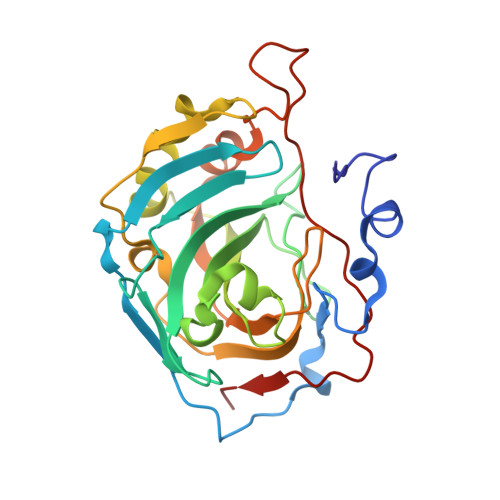
Structural and kinetic analysis of proton shuttle residues in the active site of human carbonic anhydrase III.
Elder, I., Fisher, Z., Laipis, P.J., Tu, C., McKenna, R., Silverman, D.N.(2007) Proteins 68: 337-343
- PubMed: 17427958
- DOI: https://doi.org/10.1002/prot.21403
- Primary Citation of Related Structures:
2HFW, 3UYN, 3UYQ - PubMed Abstract:
We report the X-ray crystal structures and rate constants for proton transfer in site-specific mutants of human carbonic anhydrase III (HCA III) that place a histidine residue in the active-site cavity: K64H, R67H, and K64H-R67N HCA III. Prior evidence from the exchange of 18O between CO2 and water measured by mass spectrometry shows each mutant to have enhanced proton transfer in catalysis compared with wild-type HCA III. However, His64 in K64H and K64H-R67N HCA III have at most a capacity for proton transfer that is only 13% that of His64 in HCA II. This reduced rate in mutants of HCA III is associated with a constrained side-chain conformation of His64, which is oriented outward, away from the active-site zinc in the crystal structures. This conformation appears stabilized by a prominent pi stacking interaction of the imidazole ring of His64 with the indole ring of Trp5 in mutants of HCA III. This single orientation of His64 in K64H HCA III predominates also in a double mutant K64H-R67N HCA III, indicating that the positive charge of Arg67 does not influence the observed conformation of His64 in the crystal structure. Hence, the structures and catalytic activity of these mutants of HCA III containing His64 account only in small part for the lower activity of this isozyme compared with HCA II. His67 in R67H HCA III was also shown to be a proton shuttle residue, having a capacity for proton transfer that was approximately four times that of His64 in K64H HCA III. This is most likely due to its proximity and orientation inward towards the zinc-bound solvent. These results emphasize the significance of side chain orientation and range of available conformational states as characteristics of an efficient proton shuttle in carbonic anhydrase.
Organizational Affiliation:
Department of Pharmacology and Therapeutics, University of Florida College of Medicine, Gainesville, Florida 32610-0267, USA.


