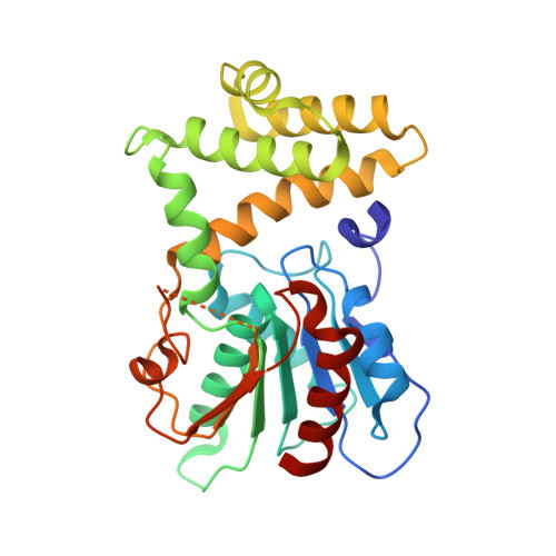
3TJM

Crystal Structure of the Human Fatty Acid Synthase Thioesterase Domain with an Activate Site-Specific Polyunsaturated Fatty Acyl Adduct


Experimental Data Snapshot

  • Method: X-RAY DIFFRACTION
  • Resolution: 1.48 Å
  • R-Value Free: 0.224 
  • R-Value Work: 0.190 
  • R-Value Observed: 0.192 

Starting Model: experimental
View more details

wwPDB Validation   3D Report Full Report


Ligand Structure Quality Assessment 


This is version 1.4 of the entry. See complete history


Literature

Crystal structure of FAS thioesterase domain with polyunsaturated fatty acyl adduct and inhibition by dihomo-gamma-linolenic acid.

Zhang, W.Chakravarty, B.Zheng, F.Gu, Z.Wu, H.Mao, J.Wakil, S.J.Quiocho, F.A.

(2011) Proc Natl Acad Sci U S A 108: 15757-15762

  • DOI: https://doi.org/10.1073/pnas.1112334108
  • Primary Citation of Related Structures:  
    3TJM

  • PubMed Abstract: 

    Human fatty acid synthase (hFAS) is a homodimeric multidomain enzyme that catalyzes a series of reactions leading to the de novo biosynthesis of long-chain fatty acids, mainly palmitate. The carboxy-terminal thioesterase (TE) domain determines the length of the fatty acyl chain and its ultimate release by hydrolysis. Because of the upregulation of hFAS in a variety of cancers, it is a target for antiproliferative agent development. Dietary long-chain polyunsaturated fatty acids (PUFAs) have been known to confer beneficial effects on many diseases and health conditions, including cancers, inflammations, diabetes, and heart diseases, but the precise molecular mechanisms involved have not been elucidated. We report the 1.48 Å crystal structure of the hFAS TE domain covalently modified and inactivated by methyl γ-linolenylfluorophosphonate. Whereas the structure confirmed the phosphorylation by the phosphonate head group of the active site serine, it also unexpectedly revealed the binding of the 18-carbon polyunsaturated γ-linolenyl tail in a long groove-tunnel site, which itself is formed mainly by the emergence of an α helix (the "helix flap"). We then found inhibition of the TE domain activity by the PUFA dihomo-γ-linolenic acid; γ- and α-linolenic acids, two popular dietary PUFAs, were less effective. Dihomo-γ-linolenic acid also inhibited fatty acid biosynthesis in 3T3-L1 preadipocytes and selective human breast cancer cell lines, including SKBR3 and MDAMB231. In addition to revealing a novel mechanism for the molecular recognition of a polyunsaturated fatty acyl chain, our results offer a new framework for developing potent FAS inhibitors as therapeutics against cancers and other diseases.


  • Organizational Affiliation

    Verna and Marrs McLean Department of Biochemistry and Molecular Biology, Baylor College of Medicine, One Baylor Plaza, Houston, TX 77030, USA.


Macromolecules
Find similar proteins by:  (by identity cutoff)  |  3D Structure
Entity ID: 1
MoleculeChains Sequence LengthOrganismDetailsImage
Fatty acid synthase283Homo sapiensMutation(s): 0 
Gene Names: FASFASN
EC: 2.3.1.85 (PDB Primary Data), 2.3.1.38 (PDB Primary Data), 2.3.1.39 (PDB Primary Data), 2.3.1.41 (PDB Primary Data), 1.1.1.100 (PDB Primary Data), 4.2.1.61 (PDB Primary Data), 1.3.1.10 (PDB Primary Data), 3.1.2.14 (PDB Primary Data), 1.3.1.39 (UniProt), 4.2.1.59 (UniProt)
UniProt & NIH Common Fund Data Resources
Find proteins for P49327 (Homo sapiens)
Explore P49327 
Go to UniProtKB:  P49327
PHAROS:  P49327
GTEx:  ENSG00000169710 
Entity Groups  
Sequence Clusters30% Identity50% Identity70% Identity90% Identity95% Identity100% Identity
UniProt GroupP49327
Sequence Annotations
Expand
  • Reference Sequence
Small Molecules
Ligands 1 Unique
IDChains Name / Formula / InChI Key2D Diagram3D Interactions
7FA
Query on 7FA

Download Ideal Coordinates CCD File 
B [auth A]methyl (R)-(6Z,9Z,12Z)-octadeca-6,9,12-trien-1-ylphosphonofluoridate
C19 H34 F O2 P
PYIHGLSRUVHCML-YMLCEXCJSA-N
Binding Affinity Annotations 
IDSourceBinding Affinity
7FA PDBBind:  3TJM Ki: 6360 (nM) from 1 assay(s)
Experimental Data & Validation

Experimental Data

  • Method: X-RAY DIFFRACTION
  • Resolution: 1.48 Å
  • R-Value Free: 0.224 
  • R-Value Work: 0.190 
  • R-Value Observed: 0.192 
  • Space Group: P 21 21 21
Unit Cell:
Length ( Å )Angle ( ˚ )
a = 50.915α = 90
b = 62.097β = 90
c = 76.511γ = 90
Software Package:
Software NamePurpose
REFMACrefinement
CNSrefinement
HKL-2000data collection
HKL-2000data reduction
SCALEPACKdata scaling
CNSphasing

Structure Validation

View Full Validation Report


Ligand Structure Quality Assessment 


Entry History 

Deposition Data

Revision History  (Full details and data files)

  • Version 1.0: 2011-09-28
    Type: Initial release
  • Version 1.1: 2011-11-16
    Changes: Database references
  • Version 1.2: 2019-07-17
    Changes: Advisory, Data collection, Derived calculations, Refinement description
  • Version 1.3: 2023-09-13
    Changes: Advisory, Data collection, Database references, Derived calculations, Refinement description
  • Version 1.4: 2024-11-06
    Changes: Structure summary