Exome sequencing identifies recurrent somatic RAC1 mutations in melanoma.
Krauthammer, M., Kong, Y., Ha, B.H., Evans, P., Bacchiocchi, A., McCusker, J.P., Cheng, E., Davis, M.J., Goh, G., Choi, M., Ariyan, S., Narayan, D., Dutton-Regester, K., Capatana, A., Holman, E.C., Bosenberg, M., Sznol, M., Kluger, H.M., Brash, D.E., Stern, D.F., Materin, M.A., Lo, R.S., Mane, S., Ma, S., Kidd, K.K., Hayward, N.K., Lifton, R.P., Schlessinger, J., Boggon, T.J., Halaban, R.(2012) Nat Genet 44: 1006-1014
- PubMed: 22842228
- DOI: https://doi.org/10.1038/ng.2359
- Primary Citation of Related Structures:
3SBD, 3SBE, 3TH5 - PubMed Abstract:
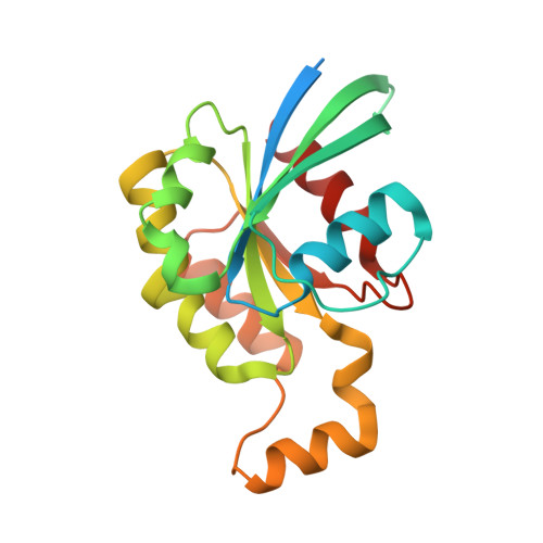
We characterized the mutational landscape of melanoma, the form of skin cancer with the highest mortality rate, by sequencing the exomes of 147 melanomas. Sun-exposed melanomas had markedly more ultraviolet (UV)-like C>T somatic mutations compared to sun-shielded acral, mucosal and uveal melanomas. Among the newly identified cancer genes was PPP6C, encoding a serine/threonine phosphatase, which harbored mutations that clustered in the active site in 12% of sun-exposed melanomas, exclusively in tumors with mutations in BRAF or NRAS. Notably, we identified a recurrent UV-signature, an activating mutation in RAC1 in 9.2% of sun-exposed melanomas. This activating mutation, the third most frequent in our cohort of sun-exposed melanoma after those of BRAF and NRAS, changes Pro29 to serine (RAC1(P29S)) in the highly conserved switch I domain. Crystal structures, and biochemical and functional studies of RAC1(P29S) showed that the alteration releases the conformational restraint conferred by the conserved proline, causes an increased binding of the protein to downstream effectors, and promotes melanocyte proliferation and migration. These findings raise the possibility that pharmacological inhibition of downstream effectors of RAC1 signaling could be of therapeutic benefit.
Organizational Affiliation:
Department of Pathology, Yale University School of Medicine, New Haven, Connecticut, USA.