Probing Mechanistic Similarities between Response Regulator Signaling Proteins and Haloacid Dehalogenase Phosphatases.
Immormino, R.M., Starbird, C.A., Silversmith, R.E., Bourret, R.B.(2015) Biochemistry 54: 3514-3527
- PubMed: 25928369
- DOI: https://doi.org/10.1021/acs.biochem.5b00286
- Primary Citation of Related Structures:
3RVJ, 3RVK, 3RVL, 3RVM, 3RVN, 3RVO, 3RVP, 3RVQ, 3RVR, 3RVS - PubMed Abstract:
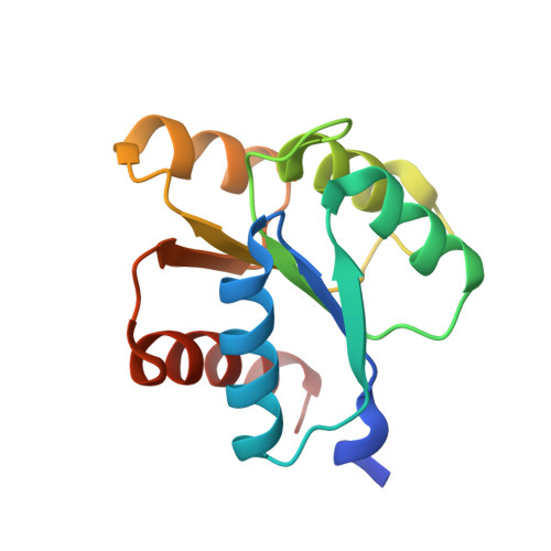
Response regulator signaling proteins and phosphatases of the haloacid dehalogenase (HAD) superfamily share strikingly similar folds, active site geometries, and reaction chemistry. Proteins from both families catalyze the transfer of a phosphoryl group from a substrate to one of their own aspartyl residues, and subsequent hydrolysis of the phosphoprotein. Notable differences include an additional Asp that functions as an acid/base catalyst and an active site well-structured prior to phosphorylation in HAD phosphatases. Both features contribute to reactions substantially faster than those for response regulators. To investigate mechanisms underlying the functional differences between response regulators and HAD phosphatases, we characterized five double mutants of the response regulator CheY designed to mimic HAD phosphatases. Each mutant contained the extra Asp paired with a phosphatase-inspired substitution to potentially position the Asp properly. Only CheY DR (Arg as the anchor) exhibited enhanced rates of both autophosphorylation with phosphoramidate and autodephosphorylation compared to those of wild-type CheY. Crystal structures of CheY DR complexed with MoO4(2-) or WO4(2-) revealed active site hydrogen bonding networks similar to those in HAD·substrate complexes, with the extra Asp positioned for direct interaction with the leaving group (phosphorylation) or nucleophile (dephosphorylation). However, CheY DR reaction kinetics did not exhibit the pH sensitivities expected for acid/base catalysis. Biochemical analysis indicated CheY DR had an enhanced propensity to adopt the active conformation without phosphorylation, but a crystal structure revealed unphosphorylated CheY DR was not locked in the active conformation. Thus, the enhanced reactivity of CheY DR reflected partial acquisition of catalytic and structural features of HAD phosphatases.
Organizational Affiliation:
Department of Microbiology and Immunology, University of North Carolina, Chapel Hill, North Carolina 27599-7290, United States.