3RNT

CRYSTAL STRUCTURE OF GUANOSINE-FREE RIBONUCLEASE T1, COMPLEXED WITH VANADATE(V), SUGGESTS CONFORMATIONAL CHANGE UPON SUBSTRATE BINDING


Experimental Data Snapshot

  • Method: X-RAY DIFFRACTION
  • Resolution: 1.80 Å
  • R-Value Observed: 0.137 

wwPDB Validation   3D Report Full Report


This is version 1.3 of the entry. See complete history


Literature

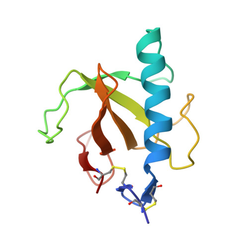
Crystal structure of guanosine-free ribonuclease T1, complexed with vanadate (V), suggests conformational change upon substrate binding.

Kostrewa, D.Choe, H.W.Heinemann, U.Saenger, W.

(1989) Biochemistry 28: 7592-7600

  • DOI: https://doi.org/10.1021/bi00445a014
  • Primary Citation of Related Structures:  
    3RNT

  • PubMed Abstract: 

    Ribonuclease T1 was crystallized in the presence of vanadate(V). The crystal structure was solved by molecular replacement and refined by least-squares methods using stereochemical restraints. The refinement was based on data between 10 and 1.8 A and converged at a crystallographic R factor of 0.137. Except for the substrate-recognition site the three-dimensional structure of ribonuclease T1 closely resembles the structure of the enzyme complexed with guanosine 2'-phosphate and its derivatives. A tetrahedral anion was found at the catalytic site and identified as H2VO4-. This is the first crystal structure of ribonuclease T1 determined in the absence of bound substrate analogue. Distinct structural differences between guanosine-free and complexed ribonuclease T1 are observed at the base-recognition site: The side chains of Tyr45 and Glu46 and the region around Asn98 changed their conformations, and the peptide bond between Asn43 and Asn44 has turned around by 140 degrees. We suggest that the structural differences seen in the crystal structures of free and complexed ribonuclease T1 are related to conformational adjustments associated with the substrate binding process.


  • Organizational Affiliation

    Institut für Kristallographie, Berlin, FRG.


Macromolecules
Find similar proteins by:  (by identity cutoff)  |  3D Structure
Entity ID: 1
MoleculeChains Sequence LengthOrganismDetailsImage
RIBONUCLEASE T1104Aspergillus oryzaeMutation(s): 0 
EC: 3.1.27.3
UniProt
Find proteins for P00651 (Aspergillus oryzae (strain ATCC 42149 / RIB 40))
Explore P00651 
Go to UniProtKB:  P00651
Entity Groups  
Sequence Clusters30% Identity50% Identity70% Identity90% Identity95% Identity100% Identity
UniProt GroupP00651
Sequence Annotations
Expand
  • Reference Sequence
Small Molecules
Ligands 2 Unique
IDChains Name / Formula / InChI Key2D Diagram3D Interactions
VO4
Query on VO4

Download Ideal Coordinates CCD File 
C [auth A]VANADATE ION
O4 V
LSGOVYNHVSXFFJ-UHFFFAOYSA-N
CA
Query on CA

Download Ideal Coordinates CCD File 
B [auth A]CALCIUM ION
Ca
BHPQYMZQTOCNFJ-UHFFFAOYSA-N
Experimental Data & Validation

Experimental Data

  • Method: X-RAY DIFFRACTION
  • Resolution: 1.80 Å
  • R-Value Observed: 0.137 
  • Space Group: P 21 21 21
Unit Cell:
Length ( Å )Angle ( ˚ )
a = 48.82α = 90
b = 46.53β = 90
c = 41.2γ = 90
Software Package:
Software NamePurpose
PROFFTrefinement

Structure Validation

View Full Validation Report


Entry History 

Deposition Data

Revision History  (Full details and data files)

  • Version 1.0: 1989-10-15
    Type: Initial release
  • Version 1.1: 2008-03-25
    Changes: Version format compliance
  • Version 1.2: 2011-07-13
    Changes: Version format compliance
  • Version 1.3: 2017-11-29
    Changes: Derived calculations, Other