3QUM

Crystal structure of human prostate specific antigen (PSA) in Fab sandwich with a high affinity and a PCa selective antibody


Experimental Data Snapshot

  • Method: X-RAY DIFFRACTION
  • Resolution: 3.20 Å
  • R-Value Free: 0.294 
  • R-Value Work: 0.198 
  • R-Value Observed: 0.203 

wwPDB Validation   3D Report Full Report


This is version 2.1 of the entry. See complete history


Literature

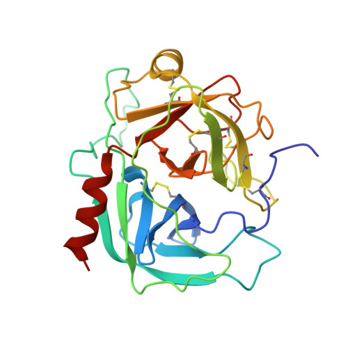
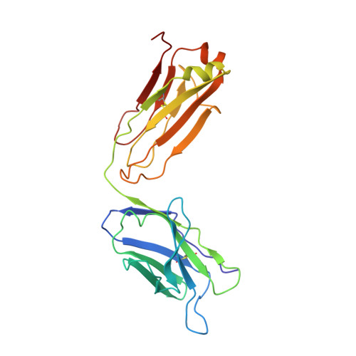
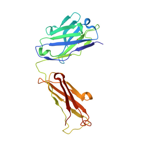
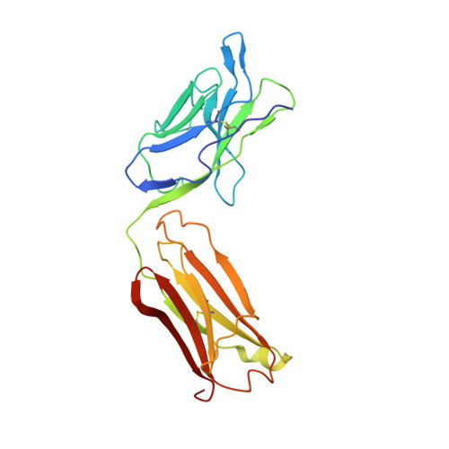
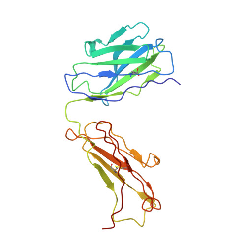
Crystal structure of human prostate-specific antigen in a sandwich antibody complex.

Stura, E.A.Muller, B.H.Bossus, M.Michel, S.Jolivet-Reynaud, C.Ducancel, F.

(2011) J Mol Biol 414: 530-544

  • DOI: https://doi.org/10.1016/j.jmb.2011.10.007
  • Primary Citation of Related Structures:  
    3QUM

  • PubMed Abstract: 

    Human prostate-specific antigen (PSA or human kallikrein-related peptidase 3) present in small quantities in the sera of healthy men becomes elevated in prostate cancer (PCa) and other prostate disorders. The ability to identify the free PSA fraction associated with PCa could increase the reliability of the PSA diagnostic test. Here we present the crystal structure of human PSA from seminal fluid in a sandwich complex with two monoclonal antibodies (mAbs). MAb 5D5A5 captures total PSA with exceptionally high affinity, and mAb 5D3D11 selectively discriminates between free PSA subforms that are more abundant in sera from patients with PCa. Although the antigen is not of seric origin, several insights into cancer diagnosis can be discerned from this complex. MAb 5D3D11 recognizes a PSA conformation different from that previously reported. Interacting with the kallikrein loop, the PSA N-linked glycan attached to asparagine 61 is an uncommonly complex sialated triantennary chain. O-linked glycosylation is observed at threonine 125. The description of how PSA subforms in prostatic fluid can be discriminated using pairs of antibodies is a first step in the design of new strategies that are capable of real discrimination among PSA subforms, which will lead to the formulation of more reliable diagnostic tests. In a companion article [Muller, B. H., Savatier, A., L'Hostis, G., Costa, N., Bossus, M., Michel, S., et al. (2011). In vitro affinity maturation of an anti-PSA antibody for prostate cancer diagnostic assay. J. Mol. Biol.], we describe engineering efforts to improve the affinity of mAb 5D3D11, a first step towards such goal.


  • Organizational Affiliation

    CEA, iBiTec-S, Service d'Ingénierie Moléculaire des Protéines, Laboratoire de Toxinologie Moléculaire et Biotechnologies, Gif-sur-Yvette F-91191, France. estura@cea.fr


Macromolecules
Find similar proteins by:  (by identity cutoff)  |  3D Structure
Entity ID: 1
MoleculeChains Sequence LengthOrganismDetailsImage
Prostate-specific antigenA [auth P],
F [auth Q]
237Homo sapiensMutation(s): 0 
EC: 3.4.21.77
UniProt & NIH Common Fund Data Resources
Find proteins for P07288 (Homo sapiens)
Explore P07288 
Go to UniProtKB:  P07288
PHAROS:  P07288
GTEx:  ENSG00000142515 
Entity Groups  
Sequence Clusters30% Identity50% Identity70% Identity90% Identity95% Identity100% Identity
UniProt GroupP07288
Sequence Annotations
Expand
  • Reference Sequence
Find similar proteins by:  (by identity cutoff)  |  3D Structure
Entity ID: 2
MoleculeChains Sequence LengthOrganismDetailsImage
Fab 5D3D11 Light ChainB [auth L],
G [auth M]
219Mus musculusMutation(s): 0 
Entity Groups  
Sequence Clusters30% Identity50% Identity70% Identity90% Identity95% Identity100% Identity
Sequence Annotations
Expand
  • Reference Sequence
Find similar proteins by:  (by identity cutoff)  |  3D Structure
Entity ID: 3
MoleculeChains Sequence LengthOrganismDetailsImage
Fab 5D3D11 Heavy ChainC [auth H],
H [auth K]
219Mus musculusMutation(s): 0 
Entity Groups  
Sequence Clusters30% Identity50% Identity70% Identity90% Identity95% Identity100% Identity
Sequence Annotations
Expand
  • Reference Sequence
Find similar proteins by:  (by identity cutoff)  |  3D Structure
Entity ID: 4
MoleculeChains Sequence LengthOrganismDetailsImage
Fab 5D5A5 Light ChainD [auth A],
I [auth C]
218Mus musculusMutation(s): 0 
Entity Groups  
Sequence Clusters30% Identity50% Identity70% Identity90% Identity95% Identity100% Identity
Sequence Annotations
Expand
  • Reference Sequence
Find similar proteins by:  (by identity cutoff)  |  3D Structure
Entity ID: 5
MoleculeChains Sequence LengthOrganismDetailsImage
Fab 5D5A5 Heavy ChainE [auth B],
J [auth D]
219Mus musculusMutation(s): 0 
Entity Groups  
Sequence Clusters30% Identity50% Identity70% Identity90% Identity95% Identity100% Identity
Sequence Annotations
Expand
  • Reference Sequence
Oligosaccharides

Help

Entity ID: 6
MoleculeChains Length2D Diagram Glycosylation3D Interactions
N-acetyl-alpha-neuraminic acid-(2-3)-beta-D-galactopyranose-(1-4)-2-acetamido-2-deoxy-beta-D-glucopyranose-(1-4)-alpha-D-mannopyranose-(1-3)-[N-acetyl-alpha-neuraminic acid-(2-3)-beta-D-galactopyranose-(1-4)-2-acetamido-2-deoxy-beta-D-glucopyranose-(1-2)-[N-acetyl-alpha-neuraminic acid-(2-3)-beta-D-galactopyranose-(1-4)-2-acetamido-2-deoxy-beta-D-glucopyranose-(1-6)]alpha-D-mannopyranose-(1-6)]alpha-D-mannopyranose-(1-4)-2-acetamido-2-deoxy-beta-D-glucopyranose-(1-4)-[alpha-L-fucopyranose-(1-6)]2-acetamido-2-deoxy-beta-D-glucopyranoseK [auth E]15N-Glycosylation
Glycosylation Resources
GlyTouCan:  G87655FC
GlyCosmos:  G87655FC
GlyGen:  G87655FC
Entity ID: 7
MoleculeChains Length2D Diagram Glycosylation3D Interactions
beta-D-galactopyranose-(1-3)-2-acetamido-2-deoxy-alpha-D-galactopyranoseL [auth F]2O-Glycosylation
Glycosylation Resources
GlyTouCan:  G00031MO
GlyCosmos:  G00031MO
GlyGen:  G00031MO
Entity ID: 8
MoleculeChains Length2D Diagram Glycosylation3D Interactions
N-acetyl-alpha-neuraminic acid-(2-3)-beta-D-galactopyranose-(1-4)-2-acetamido-2-deoxy-beta-D-glucopyranose-(1-6)-alpha-D-mannopyranose-(1-3)-[N-acetyl-alpha-neuraminic acid-(2-3)-beta-D-galactopyranose-(1-4)-2-acetamido-2-deoxy-beta-D-glucopyranose-(1-2)-[N-acetyl-alpha-neuraminic acid-(2-3)-beta-D-galactopyranose-(1-4)-2-acetamido-2-deoxy-beta-D-glucopyranose-(1-6)]alpha-D-mannopyranose-(1-6)]alpha-D-mannopyranose-(1-4)-2-acetamido-2-deoxy-beta-D-glucopyranose-(1-4)-[alpha-L-fucopyranose-(1-6)]2-acetamido-2-deoxy-beta-D-glucopyranoseM [auth G]15N-Glycosylation
Glycosylation Resources
GlyTouCan:  G67371LF
GlyCosmos:  G67371LF
GlyGen:  G67371LF
Biologically Interesting Molecules (External Reference) 1 Unique
Experimental Data & Validation

Experimental Data

  • Method: X-RAY DIFFRACTION
  • Resolution: 3.20 Å
  • R-Value Free: 0.294 
  • R-Value Work: 0.198 
  • R-Value Observed: 0.203 
  • Space Group: P 1 21 1
Unit Cell:
Length ( Å )Angle ( ˚ )
a = 55.308α = 90
b = 226.883β = 96.51
c = 118.694γ = 90
Software Package:
Software NamePurpose
ProDCdata collection
MOLREPphasing
PHENIXrefinement
MOSFLMdata reduction
SCALAdata scaling

Structure Validation

View Full Validation Report


Entry History 

Deposition Data

Revision History  (Full details and data files)

  • Version 1.0: 2011-11-09
    Type: Initial release
  • Version 1.1: 2011-12-21
    Changes: Database references
  • Version 2.0: 2020-07-29
    Type: Remediation
    Reason: Carbohydrate remediation
    Changes: Advisory, Atomic model, Data collection, Derived calculations, Structure summary
  • Version 2.1: 2023-09-13
    Changes: Advisory, Data collection, Database references, Refinement description, Structure summary