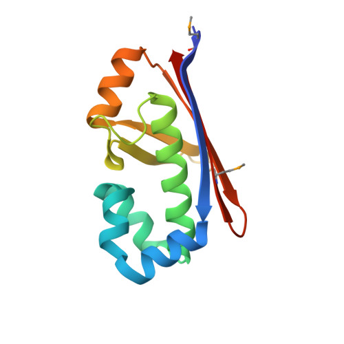
3QMN

Crystal Structure of 4'-Phosphopantetheinyl Transferase AcpS from Vibrio cholerae O1 biovar eltor


Experimental Data Snapshot

  • Method: X-RAY DIFFRACTION
  • Resolution: 1.85 Å
  • R-Value Free: 0.191 
  • R-Value Work: 0.156 
  • R-Value Observed: 0.158 

wwPDB Validation   3D Report Full Report


Ligand Structure Quality Assessment 


This is version 1.3 of the entry. See complete history


Literature

Structural characterization and comparison of three acyl-carrier-protein synthases from pathogenic bacteria.

Halavaty, A.S.Kim, Y.Minasov, G.Shuvalova, L.Dubrovska, I.Winsor, J.Zhou, M.Onopriyenko, O.Skarina, T.Papazisi, L.Kwon, K.Peterson, S.N.Joachimiak, A.Savchenko, A.Anderson, W.F.

(2012) Acta Crystallogr D Biol Crystallogr 68: 1359-1370

  • DOI: https://doi.org/10.1107/S0907444912029101
  • Primary Citation of Related Structures:  
    3HYK, 3QMN, 4JM7

  • PubMed Abstract: 

    Some bacterial type II fatty-acid synthesis (FAS II) enzymes have been shown to be important candidates for drug discovery. The scientific and medical quest for new FAS II protein targets continues to stimulate research in this field. One of the possible additional candidates is the acyl-carrier-protein synthase (AcpS) enzyme. Its holo form post-translationally modifies the apo form of an acyl carrier protein (ACP), which assures the constant delivery of thioester intermediates to the discrete enzymes of FAS II. At the Center for Structural Genomics of Infectious Diseases (CSGID), AcpSs from Staphylococcus aureus (AcpS(SA)), Vibrio cholerae (AcpS(VC)) and Bacillus anthracis (AcpS(BA)) have been structurally characterized in their apo, holo and product-bound forms, respectively. The structure of AcpS(BA) is emphasized because of the two 3',5'-adenosine diphosphate (3',5'-ADP) product molecules that are found in each of the three coenzyme A (CoA) binding sites of the trimeric protein. One 3',5'-ADP is bound as the 3',5'-ADP part of CoA in the known structures of the CoA-AcpS and 3',5'-ADP-AcpS binary complexes. The position of the second 3',5'-ADP has never been described before. It is in close proximity to the first 3',5'-ADP and the ACP-binding site. The coordination of two ADPs in AcpS(BA) may possibly be exploited for the design of AcpS inhibitors that can block binding of both CoA and ACP.


  • Organizational Affiliation

    Center for Structural Genomics of Infectious Diseases, USA.


Macromolecules
Find similar proteins by:  (by identity cutoff)  |  3D Structure
Entity ID: 1
MoleculeChains Sequence LengthOrganismDetailsImage
Holo-[acyl-carrier-protein] synthase
A, B, C, D, E
A, B, C, D, E, F, G, H, I, J, K, L, M, N, O, P, Q, R, S, T, U, V, W, X
129Vibrio cholerae O1 biovar El Tor str. N16961Mutation(s): 0 
Gene Names: acpSVC_2457
EC: 2.7.8.7
UniProt
Find proteins for Q9KPB6 (Vibrio cholerae serotype O1 (strain ATCC 39315 / El Tor Inaba N16961))
Explore Q9KPB6 
Go to UniProtKB:  Q9KPB6
Entity Groups  
Sequence Clusters30% Identity50% Identity70% Identity90% Identity95% Identity100% Identity
UniProt GroupQ9KPB6
Sequence Annotations
Expand
  • Reference Sequence
Small Molecules
Ligands 7 Unique
IDChains Name / Formula / InChI Key2D Diagram3D Interactions
COA
Query on COA

Download Ideal Coordinates CCD File 
AA [auth A]
BE [auth X]
CB [auth G]
ED [auth S]
HB [auth H]
AA [auth A],
BE [auth X],
CB [auth G],
ED [auth S],
HB [auth H],
IA [auth C],
IC [auth N],
ID [auth T],
JC [auth N],
LB [auth I],
MC [auth O],
OA [auth D],
PB [auth J],
PD [auth U],
QC [auth P],
SA [auth E],
VB [auth K],
VC [auth Q],
VD [auth W],
WD [auth W],
Y [auth A],
YA [auth F],
ZB [auth L],
ZC [auth R]
COENZYME A
C21 H36 N7 O16 P3 S
RGJOEKWQDUBAIZ-IBOSZNHHSA-N
A3P
Query on A3P

Download Ideal Coordinates CCD File 
PC [auth O]ADENOSINE-3'-5'-DIPHOSPHATE
C10 H15 N5 O10 P2
WHTCPDAXWFLDIH-KQYNXXCUSA-N
MRD
Query on MRD

Download Ideal Coordinates CCD File 
BB [auth F]
DC [auth L]
LC [auth N]
MD [auth T]
RA [auth D]
BB [auth F],
DC [auth L],
LC [auth N],
MD [auth T],
RA [auth D],
YB [auth K]
(4R)-2-METHYLPENTANE-2,4-DIOL
C6 H14 O2
SVTBMSDMJJWYQN-RXMQYKEDSA-N
MPD
Query on MPD

Download Ideal Coordinates CCD File 
CD [auth R]
DA [auth A]
DB [auth G]
FA [auth B]
FD [auth S]
CD [auth R],
DA [auth A],
DB [auth G],
FA [auth B],
FD [auth S],
HC [auth M],
IB [auth H],
MA [auth C],
NC [auth O],
OB [auth I],
OD [auth U],
RC [auth P],
SD [auth V],
VA [auth E],
XD [auth W],
YC [auth Q]
(4S)-2-METHYL-2,4-PENTANEDIOL
C6 H14 O2
SVTBMSDMJJWYQN-YFKPBYRVSA-N
ACT
Query on ACT

Download Ideal Coordinates CCD File 
AE [auth W]
DD [auth R]
EA [auth A]
LD [auth T]
NA [auth C]
AE [auth W],
DD [auth R],
EA [auth A],
LD [auth T],
NA [auth C],
UB [auth K],
XA [auth E],
XB [auth K]
ACETATE ION
C2 H3 O2
QTBSBXVTEAMEQO-UHFFFAOYSA-M
CA
Query on CA

Download Ideal Coordinates CCD File 
AB [auth F]
AC [auth L]
AD [auth R]
BA [auth A]
BC [auth L]
AB [auth F],
AC [auth L],
AD [auth R],
BA [auth A],
BC [auth L],
BD [auth R],
CA [auth A],
CE [auth X],
DE [auth X],
EB [auth G],
EC [auth M],
FB [auth G],
FC [auth M],
GA [auth B],
GD [auth S],
HA [auth B],
HD [auth S],
JA [auth C],
JB [auth H],
JD [auth T],
KA [auth C],
KB [auth H],
KC [auth N],
KD [auth T],
MB [auth I],
NB [auth I],
OC [auth O],
PA [auth D],
QA [auth D],
QB [auth J],
QD [auth U],
RD [auth U],
SB [auth J],
SC [auth P],
TA [auth E],
TB [auth K],
TC [auth P],
TD [auth V],
UA [auth E],
UD [auth V],
WB [auth K],
WC [auth Q],
XC [auth Q],
YD [auth W],
ZA [auth F],
ZD [auth W]
CALCIUM ION
Ca
BHPQYMZQTOCNFJ-UHFFFAOYSA-N
CL
Query on CL

Download Ideal Coordinates CCD File 
CC [auth L]
GB [auth G]
GC [auth M]
LA [auth C]
ND [auth T]
CC [auth L],
GB [auth G],
GC [auth M],
LA [auth C],
ND [auth T],
RB [auth J],
UC [auth P],
WA [auth E],
Z [auth A]
CHLORIDE ION
Cl
VEXZGXHMUGYJMC-UHFFFAOYSA-M
Modified Residues  1 Unique
IDChains TypeFormula2D DiagramParent
MSE
Query on MSE
A, B, C, D, E
A, B, C, D, E, F, G, H, I, J, K, L, M, N, O, P, Q, R, S, T, U, V, W, X
L-PEPTIDE LINKINGC5 H11 N O2 SeMET
Experimental Data & Validation

Experimental Data

Unit Cell:
Length ( Å )Angle ( ˚ )
a = 94.553α = 90
b = 139.039β = 93.72
c = 138.319γ = 90
Software Package:
Software NamePurpose
SBC-Collectdata collection
HKL-3000phasing
SHELXSphasing
MLPHAREphasing
RESOLVEmodel building
SOLVEphasing
PHENIXmodel building
REFMACrefinement
HKL-3000data reduction
HKL-3000data scaling
RESOLVEphasing
PHENIXphasing

Structure Validation

View Full Validation Report


Ligand Structure Quality Assessment 


Entry History 

Deposition Data

Revision History  (Full details and data files)

  • Version 1.0: 2011-03-02
    Type: Initial release
  • Version 1.1: 2011-07-13
    Changes: Version format compliance
  • Version 1.2: 2012-12-05
    Changes: Database references
  • Version 1.3: 2024-10-16
    Changes: Data collection, Database references, Derived calculations, Structure summary