3O1Y

Electron transfer complexes: Experimental mapping of the redox-dependent cytochrome c electrostatic surface


Experimental Data Snapshot

  • Method: X-RAY DIFFRACTION
  • Resolution: 1.75 Å
  • R-Value Free: 0.248 
  • R-Value Work: 0.186 
  • R-Value Observed: 0.189 

wwPDB Validation   3D Report Full Report


Ligand Structure Quality Assessment 


This is version 1.2 of the entry. See complete history


Literature

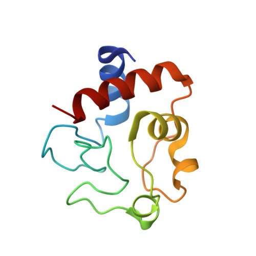
Nitrate as a probe of cytochrome c surface: crystallographic identification of crucial "hot spots" for protein-protein recognition.

De March, M.Demitri, N.De Zorzi, R.Casini, A.Gabbiani, C.Guerri, A.Messori, L.Geremia, S.

(2014) J Inorg Biochem 135: 58-67

  • DOI: https://doi.org/10.1016/j.jinorgbio.2014.02.015
  • Primary Citation of Related Structures:  
    3O1Y, 3O20

  • PubMed Abstract: 

    The electrostatic surface of cytochrome c and its changes with the iron oxidation state are involved in the docking and undocking processes of this protein to its biological partners in the mitochondrial respiratory pathway. To investigate the subtle mechanisms of formation of productive macromolecular complexes and of their breakage following the electron transfer process, the X-ray structures of horse heart ferri-cytochrome c (trigonal form) and ferro-cytochrome c (monoclinic form) were obtained using nitrate ions both as a crystallizing agent and an anionic probe for mapping the electrostatic surface changes. Both crystal forms contain three protein molecules in the asymmetric unit. In addition, a total of 21.5 and 18 crystallographically independent nitrate ions were identified for the trigonal and monoclinic forms, respectively. By matching all the six crystallographically independent protein molecules, 26 different anion-protein interaction sites were identified on the surfaces of cytochrome c, 10 of which were found in both forms, 8 present only in the oxidized and 8 only in the reduced form. The structural analysis of the electron transfer complexes, based on this new information, suggests a specific exit strategy for cytochrome c after formation of productive protein-protein complexes: a directional sliding mechanism for the electron shuttle on the surface of the redox partner is proposed to take place after the electron transfer process has occurred.


  • Organizational Affiliation

    Centre of Excellence in Biocrystallography, Department of Chemical and Pharmaceutical Sciences, University of Trieste, via L. Giorgeri 1, 34127 Trieste, Italy.


Macromolecules
Find similar proteins by:  (by identity cutoff)  |  3D Structure
Entity ID: 1
MoleculeChains Sequence LengthOrganismDetailsImage
Cytochrome c
A, B, C
105Equus caballusMutation(s): 1 
UniProt
Find proteins for P00004 (Equus caballus)
Explore P00004 
Go to UniProtKB:  P00004
Entity Groups  
Sequence Clusters30% Identity50% Identity70% Identity90% Identity95% Identity100% Identity
UniProt GroupP00004
Sequence Annotations
Expand
  • Reference Sequence
Small Molecules
Ligands 2 Unique
IDChains Name / Formula / InChI Key2D Diagram3D Interactions
HEC
Query on HEC

Download Ideal Coordinates CCD File 
D [auth A],
M [auth B],
V [auth C]
HEME C
C34 H34 Fe N4 O4
HXQIYSLZKNYNMH-LJNAALQVSA-N
NO3
Query on NO3

Download Ideal Coordinates CCD File 
AA [auth C]
BA [auth C]
E [auth A]
F [auth A]
G [auth A]
AA [auth C],
BA [auth C],
E [auth A],
F [auth A],
G [auth A],
H [auth A],
I [auth A],
J [auth A],
K [auth A],
L [auth A],
N [auth B],
O [auth B],
P [auth B],
Q [auth B],
R [auth B],
S [auth B],
T [auth B],
U [auth B],
W [auth C],
X [auth C],
Y [auth C],
Z [auth C]
NITRATE ION
N O3
NHNBFGGVMKEFGY-UHFFFAOYSA-N
Experimental Data & Validation

Experimental Data

  • Method: X-RAY DIFFRACTION
  • Resolution: 1.75 Å
  • R-Value Free: 0.248 
  • R-Value Work: 0.186 
  • R-Value Observed: 0.189 
  • Space Group: P 31 2 1
Unit Cell:
Length ( Å )Angle ( ˚ )
a = 79.695α = 90
b = 79.695β = 90
c = 89.247γ = 120
Software Package:
Software NamePurpose
AMoREphasing
REFMACrefinement
MOSFLMdata reduction
SCALAdata scaling

Structure Validation

View Full Validation Report


Ligand Structure Quality Assessment 


Entry History 

Deposition Data

Revision History  (Full details and data files)

  • Version 1.0: 2012-01-25
    Type: Initial release
  • Version 1.1: 2019-03-20
    Changes: Advisory, Data collection, Database references, Derived calculations
  • Version 1.2: 2023-09-06
    Changes: Data collection, Database references, Derived calculations, Refinement description