3NZ8

Crystal structure of the HIV-2 neutralizing Fab fragment 7C8


Experimental Data Snapshot

  • Method: X-RAY DIFFRACTION
  • Resolution: 2.70 Å
  • R-Value Free: 0.273 
  • R-Value Work: 0.226 
  • R-Value Observed: 0.229 

wwPDB Validation   3D Report Full Report


This is version 1.3 of the entry. See complete history


Literature

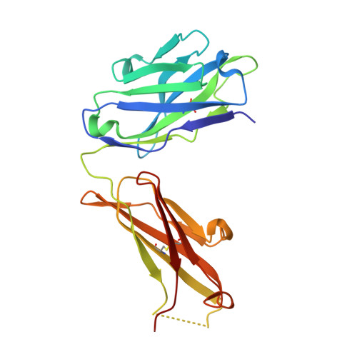
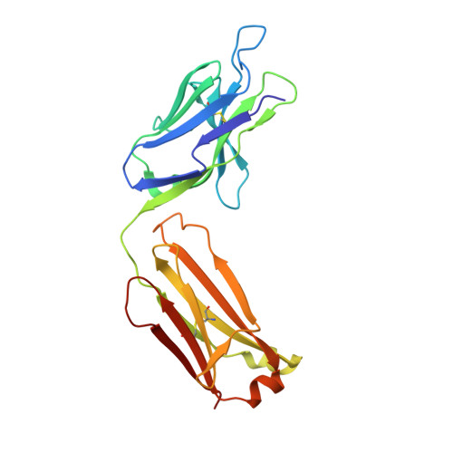
Crystal Structure of the HIV-2 Neutralizing Fab Fragment 7C8 with High Specificity to the V3 Region of gp125.

Uchtenhagen, H.Friemann, R.Raszewski, G.Spetz, A.L.Nilsson, L.Achour, A.

(2011) PLoS One 6: e18767-e18767

  • DOI: https://doi.org/10.1371/journal.pone.0018767
  • Primary Citation of Related Structures:  
    3NZ8

  • PubMed Abstract: 

    7C8 is a mouse monoclonal antibody specific for the third hypervariable region (V3) of the human immunodeficiency virus type 2 (HIV-2)-associated protein gp125. The three-dimensional crystal structure of the Fab fragment of 7C8, determined to 2.7 Å resolution, reveals a deep and narrow antigen-binding cleft with architecture appropriate for an elongated epitope. The highly hydrophobic cleft is bordered on one side by the negatively charged second complementarity determining region (CDR2) and the unusually long positively charged CDR3 of the heavy chain and, on the other side, by the CDR1 of the light chain. Analysis of 7C8 in complex with molecular models of monomeric and trimeric gp125 highlights the importance of a conserved stretch of residues FHSQ that is localized centrally on the V3 region of gp125. Furthermore, modeling also indicates that the Fab fragment neutralizes the virus by sterically impairing subsequent engagement of the gp125 trimer with the co-receptor on the target cell.


  • Organizational Affiliation

    F59 Department of Medicine, Center for Infectious Medicine, Karolinska University Hospital Huddinge, Karolinska Institutet, Stockholm, Sweden.


Macromolecules
Find similar proteins by:  (by identity cutoff)  |  3D Structure
Entity ID: 1
MoleculeChains Sequence LengthOrganismDetailsImage
Mouse anti V3 antibody 7C8 Fab, heavy chainA,
C [auth H]
221Mus musculusMutation(s): 0 
Entity Groups  
Sequence Clusters30% Identity50% Identity70% Identity90% Identity95% Identity100% Identity
Sequence Annotations
Expand
  • Reference Sequence
Find similar proteins by:  (by identity cutoff)  |  3D Structure
Entity ID: 2
MoleculeChains Sequence LengthOrganismDetailsImage
Mouse anti V3 antibody 7C8 Fab, light chainB,
D [auth L]
218Mus musculusMutation(s): 0 
Entity Groups  
Sequence Clusters30% Identity50% Identity70% Identity90% Identity95% Identity100% Identity
Sequence Annotations
Expand
  • Reference Sequence
Small Molecules
Ligands 1 Unique
IDChains Name / Formula / InChI Key2D Diagram3D Interactions
GOL
Query on GOL

Download Ideal Coordinates CCD File 
E [auth B]GLYCEROL
C3 H8 O3
PEDCQBHIVMGVHV-UHFFFAOYSA-N
Experimental Data & Validation

Experimental Data

  • Method: X-RAY DIFFRACTION
  • Resolution: 2.70 Å
  • R-Value Free: 0.273 
  • R-Value Work: 0.226 
  • R-Value Observed: 0.229 
  • Space Group: P 32 2 1
Unit Cell:
Length ( Å )Angle ( ˚ )
a = 100.071α = 90
b = 100.071β = 90
c = 196.763γ = 120
Software Package:
Software NamePurpose
MAR345dtbdata collection
PHASERphasing
REFMACrefinement
MOSFLMdata reduction
SCALAdata scaling

Structure Validation

View Full Validation Report


Entry History 

Deposition Data

Revision History  (Full details and data files)

  • Version 1.0: 2011-05-18
    Type: Initial release
  • Version 1.1: 2011-07-13
    Changes: Version format compliance
  • Version 1.2: 2018-01-24
    Changes: Advisory, Data collection, Structure summary
  • Version 1.3: 2023-09-06
    Changes: Advisory, Data collection, Database references, Derived calculations, Refinement description