3NU0

Design, Synthesis, Biological Evaluation and X-ray Crystal Structure of Novel Classical 6,5,6-TricyclicBenzo[4,5]thieno[2,3-d]pyrimidines as Dual Thymidylate Synthase and Dihydrofolate Reductase Inhibitors


Experimental Data Snapshot

  • Method: X-RAY DIFFRACTION
  • Resolution: 1.35 Å
  • R-Value Free: 0.209 
  • R-Value Work: 0.180 
  • R-Value Observed: 0.181 

wwPDB Validation   3D Report Full Report


Ligand Structure Quality Assessment 


This is version 1.3 of the entry. See complete history


Literature

Design, synthesis, biological evaluation and X-ray crystal structure of novel classical 6,5,6-tricyclic benzo[4,5]thieno[2,3-d]pyrimidines as dual thymidylate synthase and dihydrofolate reductase inhibitors.

Zhang, X.Zhou, X.Kisliuk, R.L.Piraino, J.Cody, V.Gangjee, A.

(2011) Bioorg Med Chem 19: 3585-3594

  • DOI: https://doi.org/10.1016/j.bmc.2011.03.067
  • Primary Citation of Related Structures:  
    3NTZ, 3NU0

  • PubMed Abstract: 

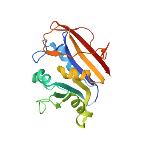
    Classical antifolates (4-7) with a tricyclic benzo[4,5]thieno[2,3-d]pyrimidine scaffold and a flexible and rigid benzoylglutamate were synthesized as dual thymidylate synthase (TS) and dihydrofolate reductase (DHFR) inhibitors. Oxidative aromatization of ethyl 2-amino-4-methyl-4,5,6,7-tetrahydro-1-benzothiophene-3-carboxylate (±)-9 to ethyl 2-amino-4-methyl-1-benzothiophene-3-carboxylate 10 with 10% Pd/C was a key synthetic step. Compounds with 2-CH₃ substituents inhibited human (h) TS (IC₅₀ =0.26-0.8 μM), but not hDHFR. Substitution of the 2-CH₃ with a 2-NH₂ increases hTS inhibition by more than 10-fold and also affords excellent hDHFR inhibition (IC₅₀ = 0.09-0.1 μM). This study shows that the tricyclic benzo[4,5]thieno[2,3-d]pyrimidine scaffold is highly conducive to single hTS or dual hTS-hDHFR inhibition depending on the 2-position substituents. The X-ray crystal structures of 6 and 7 with hDHFR reveal, for the first time, that tricyclics 6 and 7 bind with the benzo[4,5]thieno[2,3-d]pyrimidine ring in the folate binding mode with the thieno S mimicking the 4-amino of methotrexate.


  • Organizational Affiliation

    Division of Medicinal Chemistry, Graduate School of Pharmaceutical Sciences, Duquesne University, Pittsburgh, PA 15282, USA.


Macromolecules
Find similar proteins by:  (by identity cutoff)  |  3D Structure
Entity ID: 1
MoleculeChains Sequence LengthOrganismDetailsImage
Dihydrofolate reducatase186Homo sapiensMutation(s): 0 
Gene Names: DHFRDHFRP1
EC: 1.5.1.3
UniProt & NIH Common Fund Data Resources
Find proteins for P00374 (Homo sapiens)
Explore P00374 
Go to UniProtKB:  P00374
PHAROS:  P00374
GTEx:  ENSG00000228716 
Entity Groups  
Sequence Clusters30% Identity50% Identity70% Identity90% Identity95% Identity100% Identity
UniProt GroupP00374
Sequence Annotations
Expand
  • Reference Sequence
Small Molecules
Ligands 3 Unique
IDChains Name / Formula / InChI Key2D Diagram3D Interactions
NDP
Query on NDP

Download Ideal Coordinates CCD File 
B [auth A]NADPH DIHYDRO-NICOTINAMIDE-ADENINE-DINUCLEOTIDE PHOSPHATE
C21 H30 N7 O17 P3
ACFIXJIJDZMPPO-NNYOXOHSSA-N
3TU
Query on 3TU

Download Ideal Coordinates CCD File 
C [auth A](2S)-2-(5-{[(2-amino-4-oxo-3,4-dihydro[1]benzothieno[2,3-d]pyrimidin-5-yl)methyl]amino}-1-oxo-1,3-dihydro-2H-isoindol-2-yl)pentanedioic acid
C24 H21 N5 O6 S
QPDXRVRRCIFNSL-HNNXBMFYSA-N
SO4
Query on SO4

Download Ideal Coordinates CCD File 
D [auth A],
E [auth A],
F [auth A]
SULFATE ION
O4 S
QAOWNCQODCNURD-UHFFFAOYSA-L
Binding Affinity Annotations 
IDSourceBinding Affinity
3TU Binding MOAD:  3NU0 IC50: 100 (nM) from 1 assay(s)
BindingDB:  3NU0 IC50: 100 (nM) from 1 assay(s)
Experimental Data & Validation

Experimental Data

  • Method: X-RAY DIFFRACTION
  • Resolution: 1.35 Å
  • R-Value Free: 0.209 
  • R-Value Work: 0.180 
  • R-Value Observed: 0.181 
  • Space Group: H 3
Unit Cell:
Length ( Å )Angle ( ˚ )
a = 84.395α = 90
b = 84.395β = 90
c = 78.193γ = 120
Software Package:
Software NamePurpose
HKL-2000data collection
MOLREPphasing
REFMACrefinement
MOSFLMdata reduction
SCALAdata scaling

Structure Validation

View Full Validation Report


Ligand Structure Quality Assessment 


Entry History 

Deposition Data

  • Released Date: 2011-05-25 
  • Deposition Author(s): Cody, V.

Revision History  (Full details and data files)

  • Version 1.0: 2011-05-25
    Type: Initial release
  • Version 1.1: 2011-07-13
    Changes: Version format compliance
  • Version 1.2: 2017-11-08
    Changes: Refinement description
  • Version 1.3: 2024-02-21
    Changes: Data collection, Database references, Derived calculations