3NQ3

Bovine beta-lactoglobulin complex with capric acid


Experimental Data Snapshot

  • Method: X-RAY DIFFRACTION
  • Resolution: 1.90 Å
  • R-Value Free: 0.289 
  • R-Value Work: 0.238 
  • R-Value Observed: 0.241 

wwPDB Validation   3D Report Full Report


Ligand Structure Quality Assessment 


This is version 1.3 of the entry. See complete history


Literature

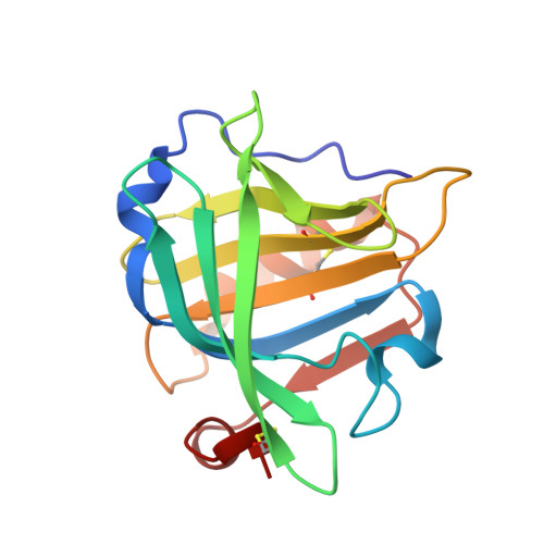
Two modes of fatty acid binding to bovine beta-lactoglobulin-crystallographic and spectroscopic studies

Loch, J.I.Polit, A.Gorecki, A.Bonarek, P.Kurpiewska, K.Dziedzicka-Wasylewska, M.Lewinski, K.

(2011) J Mol Recognit 24: 341-349

  • DOI: https://doi.org/10.1002/jmr.1084
  • Primary Citation of Related Structures:  
    3NPO, 3NQ3, 3NQ9

  • PubMed Abstract: 

    Lactoglobulin is a natural protein present in bovine milk and common component of human diet, known for binding with high affinity wide range of hydrophobic compounds, among them fatty acids 12-20 carbon atoms long. Shorter fatty acids were reported as not binding to β-lactoglobulin. We used X-ray crystallography and fluorescence spectroscopy to show that lactoglobulin binds also 8- and 10-carbon caprylic and capric acids, however with lower affinity. The determined apparent association constant for lactoglobulin complex with caprylic acid is 10.8 ± 1.7 × 10(3) M(-1), while for capric acid is 6.0 ± 0.5 × 10(3) M(-1). In crystal structures determined with resolution 1.9 Å the caprylic acid is bound in upper part of central calyx near polar residues located at CD loop, while the capric acid is buried deeper in the calyx bottom and does not interact with polar residues at CD loop. In both structures, water molecule hydrogen-bonded to carboxyl group of fatty acid is observed. Different location of ligands in the binding site indicates that competition between polar and hydrophobic interactions is an important factor determining position of the ligand in β-barrel.


  • Organizational Affiliation

    Faculty of Chemistry, Department of Crystal Physics and Crystal Chemistry, Jagiellonian University, Ingardena 3, 30-060 Kraków, Poland.


Macromolecules
Find similar proteins by:  (by identity cutoff)  |  3D Structure
Entity ID: 1
MoleculeChains Sequence LengthOrganismDetailsImage
Beta-lactoglobulin162Bos taurusMutation(s): 0 
UniProt
Find proteins for P02754 (Bos taurus)
Explore P02754 
Go to UniProtKB:  P02754
Entity Groups  
Sequence Clusters30% Identity50% Identity70% Identity90% Identity95% Identity100% Identity
UniProt GroupP02754
Sequence Annotations
Expand
  • Reference Sequence
Small Molecules
Binding Affinity Annotations 
IDSourceBinding Affinity
DKA PDBBind:  3NQ3 Kd: 1.67e+5 (nM) from 1 assay(s)
Binding MOAD:  3NQ3 Ka: 6000 (M^-1) from 1 assay(s)
Experimental Data & Validation

Experimental Data

  • Method: X-RAY DIFFRACTION
  • Resolution: 1.90 Å
  • R-Value Free: 0.289 
  • R-Value Work: 0.238 
  • R-Value Observed: 0.241 
  • Space Group: P 32 2 1
Unit Cell:
Length ( Å )Angle ( ˚ )
a = 53.5α = 90
b = 53.5β = 90
c = 112.14γ = 120
Software Package:
Software NamePurpose
CrysalisProdata collection
MOLREPphasing
REFMACrefinement
CrysalisProdata reduction
SCALAdata scaling

Structure Validation

View Full Validation Report


Ligand Structure Quality Assessment 


Entry History 

Deposition Data

Revision History  (Full details and data files)

  • Version 1.0: 2010-07-14
    Type: Initial release
  • Version 1.1: 2011-07-13
    Changes: Version format compliance
  • Version 1.2: 2023-09-06
    Changes: Data collection, Database references, Derived calculations, Structure summary
  • Version 1.3: 2023-11-01
    Changes: Refinement description