3L3R

Structural, Computational and Kinetic Data for Antifolate Interactions Against Pneumocystis jirovecii, Pneumocystis carinii and Human Dihydrofolate Reductase and Their Active Site Mutants


Experimental Data Snapshot

  • Method: X-RAY DIFFRACTION
  • Resolution: 2.00 Å
  • R-Value Free: 0.257 
  • R-Value Work: 0.171 
  • R-Value Observed: 0.175 

wwPDB Validation   3D Report Full Report


Ligand Structure Quality Assessment 


This is version 1.5 of the entry. See complete history


Literature

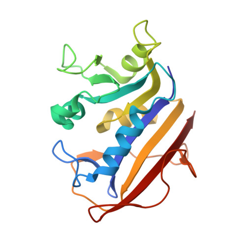
Kinetic and Structural Analysis for Potent Antifolate Inhibition of Pneumocystis jirovecii, Pneumocystis carinii, and Human Dihydrofolate Reductases and Their Active-Site Variants.

Cody, V.Pace, J.Queener, S.F.Adair, O.O.Gangjee, A.

(2013) Antimicrob Agents Chemother 57: 2669-2677

  • DOI: https://doi.org/10.1128/AAC.00172-13
  • Primary Citation of Related Structures:  
    3L3R, 3OAF, 4G8Z, 4G95

  • PubMed Abstract: 

    A major concern of immunocompromised patients, in particular those with AIDS, is susceptibility to infection caused by opportunistic pathogens such as Pneumocystis jirovecii, which is a leading cause of pneumonia in immunocompromised patients. We report the first kinetic and structural data for 2,4-diamino-6-[(2',5'-dichloro anilino)methyl]pyrido[2,3-d]pyrimidine (OAAG324), a potent inhibitor of dihydrofolate reductase (DHFR) from P. jirovecii (pjDHFR), and also for trimethoprim (TMP) and methotrexate (MTX) with pjDHFR, Pneumocystis carinii DHFR (pcDHFR), and human DHFR (hDHFR). OAAG324 shows a 9.0-fold selectivity for pjDHFR (Ki, 2.7 nM) compared to its selectivity for hDHFR (Ki, 24.4 nM), whereas there is only a 2.3-fold selectivity for pcDHFR (Ki, 6.3 nM). In order to understand the determinants of inhibitory potency, active-site mutations of pj-, pc-, and hDHFR were explored to make these enzymes more like each other. The most unexpected observations were that the variant pcDHFR forms with K37Q and K37Q/F69N mutations, which made the enzyme more like the human form, also made these enzymes more sensitive to the inhibitory activity of OAAG324, with Ki values of 0.26 and 0.71 nM, respectively. A similar gain in sensitivity was also observed for the hDHFR N64F variant, which showed a lower Ki value (0.58 nM) than native hDHFR, pcDHFR, or pjDHFR. Structural data are reported for complexes of OAAG324 with hDHFR and its Q35K and Q35S/N64F variants and for the complex of the K37S/F69N variant of pcDHFR with TMP. These results provide useful insight into the role of these residues in the optimization of highly selective inhibitors of DHFR against the opportunistic pathogen P. jirovecii.


  • Organizational Affiliation

    Structural Biology Department, Hauptman Woodward Medical Research Institute, Buffalo, New York, USA.


Macromolecules
Find similar proteins by:  (by identity cutoff)  |  3D Structure
Entity ID: 1
MoleculeChains Sequence LengthOrganismDetailsImage
Dihydrofolate reductase186Homo sapiensMutation(s): 1 
EC: 1.5.1.3
UniProt & NIH Common Fund Data Resources
Find proteins for P00374 (Homo sapiens)
Explore P00374 
Go to UniProtKB:  P00374
PHAROS:  P00374
GTEx:  ENSG00000228716 
Entity Groups  
Sequence Clusters30% Identity50% Identity70% Identity90% Identity95% Identity100% Identity
UniProt GroupP00374
Sequence Annotations
Expand
  • Reference Sequence
Small Molecules
Ligands 3 Unique
IDChains Name / Formula / InChI Key2D Diagram3D Interactions
NDP
Query on NDP

Download Ideal Coordinates CCD File 
C [auth A]NADPH DIHYDRO-NICOTINAMIDE-ADENINE-DINUCLEOTIDE PHOSPHATE
C21 H30 N7 O17 P3
ACFIXJIJDZMPPO-NNYOXOHSSA-N
OAG
Query on OAG

Download Ideal Coordinates CCD File 
B [auth A]6-{[(2,5-dichlorophenyl)amino]methyl}pyrido[2,3-d]pyrimidine-2,4-diamine
C14 H12 Cl2 N6
RSPDQFOJWPXTPN-UHFFFAOYSA-N
SO4
Query on SO4

Download Ideal Coordinates CCD File 
D [auth A]SULFATE ION
O4 S
QAOWNCQODCNURD-UHFFFAOYSA-L
Binding Affinity Annotations 
IDSourceBinding Affinity
OAG BindingDB:  3L3R Ki: 24 (nM) from 1 assay(s)
IC50: 6900 (nM) from 1 assay(s)
Experimental Data & Validation

Experimental Data

  • Method: X-RAY DIFFRACTION
  • Resolution: 2.00 Å
  • R-Value Free: 0.257 
  • R-Value Work: 0.171 
  • R-Value Observed: 0.175 
  • Space Group: H 3
Unit Cell:
Length ( Å )Angle ( ˚ )
a = 84.53α = 90
b = 84.53β = 90
c = 78.423γ = 120
Software Package:
Software NamePurpose
CrystalCleardata collection
MOLREPphasing
REFMACrefinement
MOSFLMdata reduction
SCALAdata scaling

Structure Validation

View Full Validation Report


Ligand Structure Quality Assessment 


Entry History 

Deposition Data

  • Released Date: 2011-01-05 
  • Deposition Author(s): Cody, V.

Revision History  (Full details and data files)

  • Version 1.0: 2011-01-05
    Type: Initial release
  • Version 1.1: 2011-07-13
    Changes: Version format compliance
  • Version 1.2: 2013-05-29
    Changes: Database references
  • Version 1.3: 2017-11-01
    Changes: Refinement description
  • Version 1.4: 2021-10-13
    Changes: Database references, Derived calculations
  • Version 1.5: 2023-09-06
    Changes: Data collection, Refinement description