3KEJ

Crystal Structure of Human MMP-13 complexed with a (pyridin-4-yl)-2H-tetrazole compound


Experimental Data Snapshot

  • Method: X-RAY DIFFRACTION
  • Resolution: 2.30 Å
  • R-Value Free: 0.236 
  • R-Value Work: 0.183 
  • R-Value Observed: 0.186 

wwPDB Validation   3D Report Full Report


Ligand Structure Quality Assessment 


This is version 1.2 of the entry. See complete history


Literature

Discovery of (pyridin-4-yl)-2H-tetrazole as a novel scaffold to identify highly selective matrix metalloproteinase-13 inhibitors for the treatment of osteoarthritis.

Schnute, M.E.O'Brien, P.M.Nahra, J.Morris, M.Howard Roark, W.Hanau, C.E.Ruminski, P.G.Scholten, J.A.Fletcher, T.R.Hamper, B.C.Carroll, J.N.Patt, W.C.Shieh, H.S.Collins, B.Pavlovsky, A.G.Palmquist, K.E.Aston, K.W.Hitchcock, J.Rogers, M.D.McDonald, J.Johnson, A.R.Munie, G.E.Wittwer, A.J.Man, C.F.Settle, S.L.Nemirovskiy, O.Vickery, L.E.Agawal, A.Dyer, R.D.Sunyer, T.

(2010) Bioorg Med Chem Lett 20: 576-580

  • DOI: https://doi.org/10.1016/j.bmcl.2009.11.081
  • Primary Citation of Related Structures:  
    3KEC, 3KEJ, 3KEK

  • PubMed Abstract: 

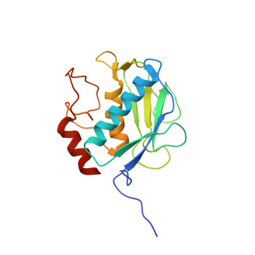
    Potent, highly selective and orally-bioavailable MMP-13 inhibitors have been identified based upon a (pyridin-4-yl)-2H-tetrazole scaffold. Co-crystal structure analysis revealed that the inhibitors bind at the S(1)(') active site pocket and are not ligands for the catalytic zinc atom. Compound 29b demonstrated reduction of cartilage degradation biomarker (TIINE) levels associated with cartilage protection in a preclinical rat osteoarthritis model.


  • Organizational Affiliation

    Global Research and Development, Pfizer Inc., 700 Chesterfield Parkway West, St. Louis, MO 63017, USA. mark.e.schnute@pfizer.com


Macromolecules
Find similar proteins by:  (by identity cutoff)  |  3D Structure
Entity ID: 1
MoleculeChains Sequence LengthOrganismDetailsImage
Collagenase 3
A, B
167Homo sapiensMutation(s): 0 
Gene Names: MMP13
EC: 3.4.24
UniProt & NIH Common Fund Data Resources
Find proteins for P45452 (Homo sapiens)
Explore P45452 
Go to UniProtKB:  P45452
PHAROS:  P45452
GTEx:  ENSG00000137745 
Entity Groups  
Sequence Clusters30% Identity50% Identity70% Identity90% Identity95% Identity100% Identity
UniProt GroupP45452
Sequence Annotations
Expand
  • Reference Sequence
Small Molecules
Ligands 3 Unique
IDChains Name / Formula / InChI Key2D Diagram3D Interactions
3EJ
Query on 3EJ

Download Ideal Coordinates CCD File 
H [auth A],
N [auth B]
4-[(5-{2-[(3-fluorobenzyl)carbamoyl]pyridin-4-yl}-2H-tetrazol-2-yl)methyl]benzoic acid
C22 H17 F N6 O3
VMAHZFWSXKJIIT-UHFFFAOYSA-N
ZN
Query on ZN

Download Ideal Coordinates CCD File 
C [auth A],
D [auth A],
I [auth B],
J [auth B]
ZINC ION
Zn
PTFCDOFLOPIGGS-UHFFFAOYSA-N
CA
Query on CA

Download Ideal Coordinates CCD File 
E [auth A]
F [auth A]
G [auth A]
K [auth B]
L [auth B]
E [auth A],
F [auth A],
G [auth A],
K [auth B],
L [auth B],
M [auth B]
CALCIUM ION
Ca
BHPQYMZQTOCNFJ-UHFFFAOYSA-N
Binding Affinity Annotations 
IDSourceBinding Affinity
3EJ PDBBind:  3KEJ Ki: 75.9 (nM) from 1 assay(s)
BindingDB:  3KEJ Ki: 76 (nM) from 1 assay(s)
Binding MOAD:  3KEJ Ki: 75.9 (nM) from 1 assay(s)
Experimental Data & Validation

Experimental Data

  • Method: X-RAY DIFFRACTION
  • Resolution: 2.30 Å
  • R-Value Free: 0.236 
  • R-Value Work: 0.183 
  • R-Value Observed: 0.186 
  • Space Group: P 21 21 2
Unit Cell:
Length ( Å )Angle ( ˚ )
a = 120.883α = 90
b = 96.457β = 90
c = 36.638γ = 90
Software Package:
Software NamePurpose
HKL-2000data collection
MOLREPphasing
REFMACrefinement
HKL-2000data reduction
HKL-2000data scaling

Structure Validation

View Full Validation Report


Ligand Structure Quality Assessment 


Entry History 

Deposition Data

Revision History  (Full details and data files)

  • Version 1.0: 2010-11-10
    Type: Initial release
  • Version 1.1: 2011-07-13
    Changes: Version format compliance
  • Version 1.2: 2024-02-21
    Changes: Data collection, Database references, Derived calculations