3K3B

Co-crystal structure of the human kinesin Eg5 with a novel tetrahydro-beta-carboline


Experimental Data Snapshot

  • Method: X-RAY DIFFRACTION
  • Resolution: 2.40 Å
  • R-Value Free: 0.243 
  • R-Value Work: 0.205 
  • R-Value Observed: 0.207 

wwPDB Validation   3D Report Full Report


Ligand Structure Quality Assessment 


This is version 1.3 of the entry. See complete history


Literature

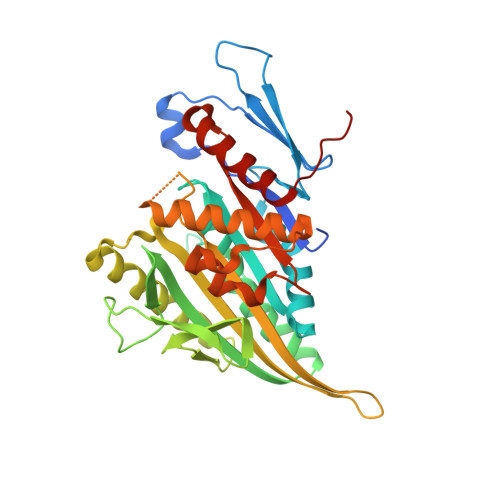
The discovery of tetrahydro-beta-carbolines as inhibitors of the kinesin Eg5.

Barsanti, P.A.Wang, W.Ni, Z.J.Duhl, D.Brammeier, N.Martin, E.Bussiere, D.Walter, A.O.

(2010) Bioorg Med Chem Lett 20: 157-160

  • DOI: https://doi.org/10.1016/j.bmcl.2009.11.012
  • Primary Citation of Related Structures:  
    3K3B

  • PubMed Abstract: 

    A series of tetrahydro-beta-carbolines were identified by HTS as inhibitors of the kinesin Eg5. Molecular modeling and medicinal chemistry techniques were employed to explore the SAR for this series with a focus of removing potential metabolic liabilities and improving cellular potency.


  • Organizational Affiliation

    Global Discovery Chemistry/Oncology and Exploratory Chemistry, Novartis Institutes for Biomedical Research, 5400 Hollis Street, Emeryville, CA 94608, USA. paul.barsanti@novartis.com


Macromolecules
Find similar proteins by:  (by identity cutoff)  |  3D Structure
Entity ID: 1
MoleculeChains Sequence LengthOrganismDetailsImage
Kinesin-like protein KIF11
A, B
368Homo sapiensMutation(s): 0 
Gene Names: KIF11EG5KNSL1TRIP5
UniProt & NIH Common Fund Data Resources
Find proteins for P52732 (Homo sapiens)
Explore P52732 
Go to UniProtKB:  P52732
PHAROS:  P52732
GTEx:  ENSG00000138160 
Entity Groups  
Sequence Clusters30% Identity50% Identity70% Identity90% Identity95% Identity100% Identity
UniProt GroupP52732
Sequence Annotations
Expand
  • Reference Sequence
Small Molecules
Ligands 6 Unique
IDChains Name / Formula / InChI Key2D Diagram3D Interactions
ADP
Query on ADP

Download Ideal Coordinates CCD File 
D [auth A],
K [auth B]
ADENOSINE-5'-DIPHOSPHATE
C10 H15 N5 O10 P2
XTWYTFMLZFPYCI-KQYNXXCUSA-N
L31
Query on L31

Download Ideal Coordinates CCD File 
E [auth A],
L [auth B]
3-[(1R)-2-acetyl-6-methyl-2,3,4,9-tetrahydro-1H-beta-carbolin-1-yl]phenol
C20 H20 N2 O2
ZNGGYWGHJZJJDR-HXUWFJFHSA-N
PEG
Query on PEG

Download Ideal Coordinates CCD File 
H [auth A]DI(HYDROXYETHYL)ETHER
C4 H10 O3
MTHSVFCYNBDYFN-UHFFFAOYSA-N
NO3
Query on NO3

Download Ideal Coordinates CCD File 
F [auth A],
I [auth B]
NITRATE ION
N O3
NHNBFGGVMKEFGY-UHFFFAOYSA-N
CL
Query on CL

Download Ideal Coordinates CCD File 
G [auth A]CHLORIDE ION
Cl
VEXZGXHMUGYJMC-UHFFFAOYSA-M
MG
Query on MG

Download Ideal Coordinates CCD File 
C [auth A],
J [auth B]
MAGNESIUM ION
Mg
JLVVSXFLKOJNIY-UHFFFAOYSA-N
Binding Affinity Annotations 
IDSourceBinding Affinity
L31 BindingDB:  3K3B IC50: 200 (nM) from 1 assay(s)
PDBBind:  3K3B IC50: 200 (nM) from 1 assay(s)
Binding MOAD:  3K3B IC50: 200 (nM) from 1 assay(s)
Experimental Data & Validation

Experimental Data

  • Method: X-RAY DIFFRACTION
  • Resolution: 2.40 Å
  • R-Value Free: 0.243 
  • R-Value Work: 0.205 
  • R-Value Observed: 0.207 
  • Space Group: P 21 21 21
Unit Cell:
Length ( Å )Angle ( ˚ )
a = 78.97α = 90
b = 92.67β = 90
c = 102.95γ = 90
Software Package:
Software NamePurpose
Blu-Icedata collection
EPMRphasing
PHENIXrefinement
MOSFLMdata reduction
SCALAdata scaling

Structure Validation

View Full Validation Report


Ligand Structure Quality Assessment 


Entry History 

Deposition Data

Revision History  (Full details and data files)

  • Version 1.0: 2009-12-15
    Type: Initial release
  • Version 1.1: 2011-07-13
    Changes: Version format compliance
  • Version 1.2: 2013-02-13
    Changes: Refinement description
  • Version 1.3: 2024-02-21
    Changes: Data collection, Database references, Derived calculations