3J1U

Low affinity dynein microtubule binding domain - tubulin complex


Experimental Data Snapshot

  • Method: ELECTRON MICROSCOPY
  • Resolution: 9.70 Å
  • Aggregation State: FILAMENT 
  • Reconstruction Method: HELICAL 

wwPDB Validation   3D Report Full Report


This is version 1.4 of the entry. See complete history


Literature

Structural basis for microtubule binding and release by dynein.

Redwine, W.B.Hernandez-Lopez, R.Zou, S.Huang, J.Reck-Peterson, S.L.Leschziner, A.E.

(2012) Science 337: 1532-1536

  • DOI: https://doi.org/10.1126/science.1224151
  • Primary Citation of Related Structures:  
    3J1T, 3J1U

  • PubMed Abstract: 

    Cytoplasmic dynein is a microtubule-based motor required for intracellular transport and cell division. Its movement involves coupling cycles of track binding and release with cycles of force-generating nucleotide hydrolysis. How this is accomplished given the ~25 nanometers separating dynein's track- and nucleotide-binding sites is not understood. Here, we present a subnanometer-resolution structure of dynein's microtubule-binding domain bound to microtubules by cryo-electron microscopy that was used to generate a pseudo-atomic model of the complex with molecular dynamics. We identified large rearrangements triggered by track binding and specific interactions, confirmed by mutagenesis and single-molecule motility assays, which tune dynein's affinity for microtubules. Our results provide a molecular model for how dynein's binding to microtubules is communicated to the rest of the motor.


  • Organizational Affiliation

    Department of Molecular and Cellular Biology, Harvard University, 52 Oxford Street, Cambridge, MA 02138, United States.


Macromolecules
Find similar proteins by:  (by identity cutoff)  |  3D Structure
Entity ID: 1
MoleculeChains Sequence LengthOrganismDetailsImage
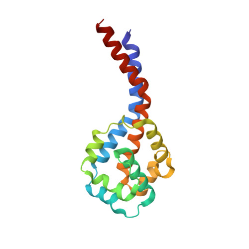
Cytoplasmic dynein 1 heavy chain 1, seryl t-RNA synthetase chimera164Mus musculusMutation(s): 0 
Gene Names: Dync1h1Dhc1Dnch1Dnchc1Dyhc
UniProt & NIH Common Fund Data Resources
Find proteins for Q9JHU4 (Mus musculus)
Explore Q9JHU4 
Go to UniProtKB:  Q9JHU4
IMPC:  MGI:103147
Entity Groups  
Sequence Clusters30% Identity50% Identity70% Identity90% Identity95% Identity100% Identity
UniProt GroupQ9JHU4
Sequence Annotations
Expand
  • Reference Sequence
Find similar proteins by:  (by identity cutoff)  |  3D Structure
Entity ID: 2
MoleculeChains Sequence LengthOrganismDetailsImage
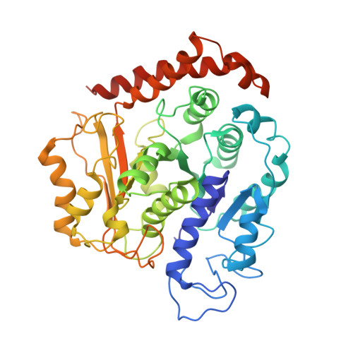
Tubulin alpha-1B chain451Bos taurusMutation(s): 0 
UniProt
Find proteins for P81947 (Bos taurus)
Explore P81947 
Go to UniProtKB:  P81947
Entity Groups  
Sequence Clusters30% Identity50% Identity70% Identity90% Identity95% Identity100% Identity
UniProt GroupP81947
Sequence Annotations
Expand
  • Reference Sequence
Find similar proteins by:  (by identity cutoff)  |  3D Structure
Entity ID: 3
MoleculeChains Sequence LengthOrganismDetailsImage
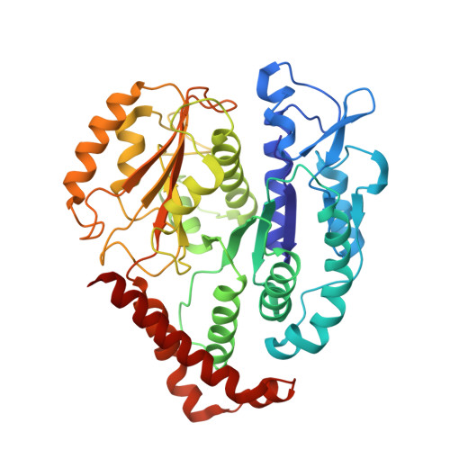
Tubulin beta-2B chain427Bos taurusMutation(s): 0 
UniProt
Find proteins for Q6B856 (Bos taurus)
Explore Q6B856 
Go to UniProtKB:  Q6B856
Entity Groups  
Sequence Clusters30% Identity50% Identity70% Identity90% Identity95% Identity100% Identity
UniProt GroupQ6B856
Sequence Annotations
Expand
  • Reference Sequence
Experimental Data & Validation

Experimental Data

  • Method: ELECTRON MICROSCOPY
  • Resolution: 9.70 Å
  • Aggregation State: FILAMENT 
  • Reconstruction Method: HELICAL 
EM Software:
TaskSoftware PackageVersion
RECONSTRUCTIONFREALIGN
RECONSTRUCTIONSPIDER

Structure Validation

View Full Validation Report


Entry History 

Deposition Data

Revision History  (Full details and data files)

  • Version 1.0: 2012-09-26
    Type: Initial release
  • Version 1.1: 2012-10-03
    Changes: Database references
  • Version 1.2: 2014-08-27
    Changes: Other
  • Version 1.3: 2018-07-18
    Changes: Author supporting evidence, Data collection
  • Version 1.4: 2024-02-21
    Changes: Data collection, Database references, Refinement description