3J0S

Remodeling of actin filaments by ADF cofilin proteins


Experimental Data Snapshot

  • Method: ELECTRON MICROSCOPY
  • Resolution: 9.00 Å
  • Aggregation State: FILAMENT 
  • Reconstruction Method: HELICAL 

wwPDB Validation   3D Report Full Report


This is version 1.4 of the entry. See complete history


Literature

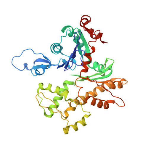
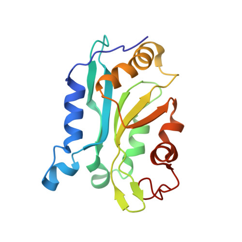
Remodeling of actin filaments by ADF/cofilin proteins.

Galkin, V.E.Orlova, A.Kudryashov, D.S.Solodukhin, A.Reisler, E.Schroder, G.F.Egelman, E.H.

(2011) Proc Natl Acad Sci U S A 108: 20568-20572

  • DOI: https://doi.org/10.1073/pnas.1110109108
  • Primary Citation of Related Structures:  
    3J0S

  • PubMed Abstract: 

    Cofilin/ADF proteins play key roles in the dynamics of actin, one of the most abundant and highly conserved eukaryotic proteins. We used cryoelectron microscopy to generate a 9-Å resolution three-dimensional reconstruction of cofilin-decorated actin filaments, the highest resolution achieved for a complex of F-actin with an actin-binding protein. We show that the cofilin-induced change in the filament twist is due to a unique conformation of the actin molecule unrelated to any previously observed state. The changes between the actin protomer in naked F-actin and in the actin-cofilin filament are greater than the conformational changes between G- and F-actin. Our results show the structural plasticity of actin, suggest that other actin-binding proteins may also induce large but different conformational changes, and show that F-actin cannot be described by a single molecular model.


  • Organizational Affiliation

    Department of Biochemistry and Molecular Genetics, University of Virginia, Box 800733, Charlottesville, VA 22908-0733, USA. galkin@virginia.edu


Macromolecules
Find similar proteins by:  (by identity cutoff)  |  3D Structure
Entity ID: 1
MoleculeChains Sequence LengthOrganismDetailsImage
Actin, cytoplasmic 1
A, B, C, D, E
A, B, C, D, E, F, G, H, I, J, K, L
374Gallus gallusMutation(s): 0 
UniProt
Find proteins for P60706 (Gallus gallus)
Explore P60706 
Go to UniProtKB:  P60706
Entity Groups  
Sequence Clusters30% Identity50% Identity70% Identity90% Identity95% Identity100% Identity
UniProt GroupP60706
Sequence Annotations
Expand
  • Reference Sequence
Find similar proteins by:  (by identity cutoff)  |  3D Structure
Entity ID: 2
MoleculeChains Sequence LengthOrganismDetailsImage
Cofilin-2166Homo sapiensMutation(s): 0 
Gene Names: CFL1CFL
UniProt & NIH Common Fund Data Resources
Find proteins for P23528 (Homo sapiens)
Explore P23528 
Go to UniProtKB:  P23528
PHAROS:  P23528
GTEx:  ENSG00000172757 
Entity Groups  
Sequence Clusters30% Identity50% Identity70% Identity90% Identity95% Identity100% Identity
UniProt GroupP23528
Sequence Annotations
Expand
  • Reference Sequence
Experimental Data & Validation

Experimental Data

  • Method: ELECTRON MICROSCOPY
  • Resolution: 9.00 Å
  • Aggregation State: FILAMENT 
  • Reconstruction Method: HELICAL 
EM Software:
TaskSoftware PackageVersion
RECONSTRUCTIONIHRSR
RECONSTRUCTIONSPIDER

Structure Validation

View Full Validation Report


Entry History 

Deposition Data

Revision History  (Full details and data files)

  • Version 1.0: 2011-12-21
    Type: Initial release
  • Version 1.1: 2012-01-11
    Changes: Database references
  • Version 1.2: 2012-06-06
    Changes: Database references
  • Version 1.3: 2018-07-18
    Changes: Author supporting evidence, Data collection
  • Version 1.4: 2024-02-21
    Changes: Data collection, Database references