


Parsing the functional specificity of Siderocalin / Lipocalin 2 / NGAL for siderophores and related small-molecule ligands
Clifton, M.C., Rupert, P.B., Hoette, T.M., Raymond, K.N., Abergel, R.J., Strong, R.K.To be published.
Experimental Data Snapshot
Entity ID: 1 | |||||
|---|---|---|---|---|---|
| Molecule | Chains | Sequence Length | Organism | Details | Image |
| Neutrophil gelatinase-associated lipocalin | 198 | Homo sapiens | Mutation(s): 2 Gene Names: HNL, LCN2, NGAL | 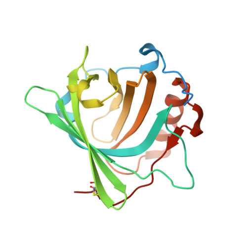 | |
UniProt & NIH Common Fund Data Resources | |||||
Find proteins for P80188 (Homo sapiens) Explore P80188 Go to UniProtKB: P80188 | |||||
PHAROS: P80188 GTEx: ENSG00000148346 | |||||
Entity Groups | |||||
| Sequence Clusters | 30% Identity50% Identity70% Identity90% Identity95% Identity100% Identity | ||||
| UniProt Group | P80188 | ||||
Sequence AnnotationsExpand | |||||
| |||||
| Ligands 7 Unique | |||||
|---|---|---|---|---|---|
| ID | Chains | Name / Formula / InChI Key | 2D Diagram | 3D Interactions | |
| MCJ Query on MCJ | N [auth C] | N-[(2,3-dihydroxyphenyl)carbonyl]-O-[(2S)-2-{[(2,3-dihydroxyphenyl)carbonyl]amino}-3-({N-[(2,3-dihydroxyphenyl)carbonyl]-L-seryl}oxy)propanoyl]-D-serine C30 H29 N3 O16 NTWRWGRCGVKQNS-KSZLIROESA-N |  | ||
| 2DS Query on 2DS | G [auth A] | N-[(2,3-dihydroxyphenyl)carbonyl]-O-[(2R)-2-{[(2,3-dihydroxyphenyl)carbonyl]amino}-3-hydroxypropanoyl]-L-serine C20 H20 N2 O11 KLXJDVFEFZPIMN-NEPJUHHUSA-N |  | ||
| 3ET Query on 3ET | F [auth A] | O-[(2R)-2-amino-3-(D-seryloxy)propanoyl]-N-[(2,3-dihydroxyphenyl)carbonyl]-L-serine C16 H21 N3 O10 ZXSIADNPWRCRTI-BBBLOLIVSA-N |  | ||
| DBH Query on DBH | I [auth B] | 2,3-DIHYDROXY-BENZOIC ACID C7 H6 O4 GLDQAMYCGOIJDV-UHFFFAOYSA-N |  | ||
| SO4 Query on SO4 | D [auth A], J [auth C] | SULFATE ION O4 S QAOWNCQODCNURD-UHFFFAOYSA-L |  | ||
| GOL Query on GOL | L [auth C], M [auth C] | GLYCEROL C3 H8 O3 PEDCQBHIVMGVHV-UHFFFAOYSA-N |  | ||
| FE Query on FE | E [auth A], H [auth B], K [auth C] | FE (III) ION Fe VTLYFUHAOXGGBS-UHFFFAOYSA-N |  | ||
| Length ( Å ) | Angle ( ˚ ) |
|---|---|
| a = 114.253 | α = 90 |
| b = 114.253 | β = 90 |
| c = 117.95 | γ = 90 |
| Software Name | Purpose |
|---|---|
| HKL-2000 | data collection |
| REFMAC | refinement |
| HKL-2000 | data reduction |
| HKL-2000 | data scaling |
| REFMAC | phasing |
RCSB PDB (citation) is hosted by
RCSB PDB is a member of the

