3HIF

The crystal structure of apo wild type CAP at 3.6 A resolution.


Experimental Data Snapshot

  • Method: X-RAY DIFFRACTION
  • Resolution: 3.59 Å
  • R-Value Free: 0.318 
  • R-Value Work: 0.295 
  • R-Value Observed: 0.296 

wwPDB Validation   3D Report Full Report


This is version 1.3 of the entry. See complete history


Literature

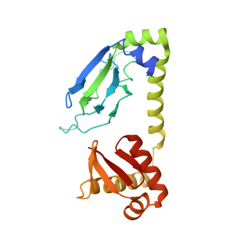
Structure of apo-CAP reveals that large conformational changes are necessary for DNA binding.

Sharma, H.Yu, S.Kong, J.Wang, J.Steitz, T.A.

(2009) Proc Natl Acad Sci U S A 106: 16604-16609

  • DOI: https://doi.org/10.1073/pnas.0908380106
  • Primary Citation of Related Structures:  
    3FWE, 3HIF

  • PubMed Abstract: 

    The binding of cAMP to the Escherichia coli catabolite gene activator protein (CAP) produces a conformational change that enables it to bind specific DNA sequences and regulate transcription, which it cannot do in the absence of the nucleotide. The crystal structures of the unliganded CAP containing a D138L mutation and the unliganded WT CAP were determined at 2.3 and 3.6 A resolution, respectively, and reveal that the two DNA binding domains have dimerized into one rigid body and their two DNA recognition helices become buried. The WT structure shows multiple orientations of this rigid body relative to the nucleotide binding domain supporting earlier biochemical data suggesting that the inactive form exists in an equilibrium among different conformations. Comparison of the structures of the liganded and unliganded CAP suggests that cAMP stabilizes the active DNA binding conformation of CAP through the interactions that the N(6) of the adenosine makes with the C-helices. These interactions are associated with the reorientation and elongation of the C-helices that precludes the formation of the inactive structure.


  • Organizational Affiliation

    Departments of Chemistry and Molecular Biophysics and Biochemistry, Yale University, Howard Hughes Medical Institute, New Haven, CT 06511, USA.


Macromolecules
Find similar proteins by:  (by identity cutoff)  |  3D Structure
Entity ID: 1
MoleculeChains Sequence LengthOrganismDetailsImage
Catabolite gene activator
A, B, C, D, E
A, B, C, D, E, F
210Escherichia coliMutation(s): 0 
Gene Names: crpS4387SF3376
UniProt
Find proteins for P0ACJ8 (Escherichia coli (strain K12))
Explore P0ACJ8 
Go to UniProtKB:  P0ACJ8
Entity Groups  
Sequence Clusters30% Identity50% Identity70% Identity90% Identity95% Identity100% Identity
UniProt GroupP0ACJ8
Sequence Annotations
Expand
  • Reference Sequence
Experimental Data & Validation

Experimental Data

  • Method: X-RAY DIFFRACTION
  • Resolution: 3.59 Å
  • R-Value Free: 0.318 
  • R-Value Work: 0.295 
  • R-Value Observed: 0.296 
  • Space Group: P 31 2 1
Unit Cell:
Length ( Å )Angle ( ˚ )
a = 125.291α = 90
b = 125.291β = 90
c = 224.677γ = 120
Software Package:
Software NamePurpose
HKL-2000data collection
DMmodel building
REFMACrefinement
HKL-2000data reduction
HKL-2000data scaling
DMphasing

Structure Validation

View Full Validation Report


Entry History 

Deposition Data

Revision History  (Full details and data files)

  • Version 1.0: 2009-09-08
    Type: Initial release
  • Version 1.1: 2011-07-13
    Changes: Advisory, Version format compliance
  • Version 1.2: 2024-02-21
    Changes: Data collection, Database references
  • Version 1.3: 2024-04-03
    Changes: Refinement description