3D6C

Crystal structure of Staphylococcal nuclease variant PHS L38E at cryogenic temperature


Experimental Data Snapshot

  • Method: X-RAY DIFFRACTION
  • Resolution: 2.00 Å
  • R-Value Free: 0.234 
  • R-Value Work: 0.173 
  • R-Value Observed: 0.179 

wwPDB Validation   3D Report Full Report


Ligand Structure Quality Assessment 


This is version 1.5 of the entry. See complete history


Literature

The pK(a) values of acidic and basic residues buried at the same internal location in a protein are governed by different factors.

Harms, M.J.Castaneda, C.A.Schlessman, J.L.Sue, G.R.Isom, D.G.Cannon, B.R.Garcia-Moreno, E.B.

(2009) J Mol Biol 389: 34-47

  • DOI: https://doi.org/10.1016/j.jmb.2009.03.039
  • Primary Citation of Related Structures:  
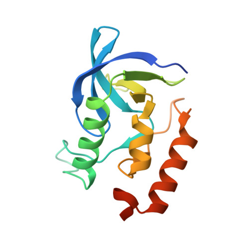
    3D6C

  • PubMed Abstract: 

    The pK(a) values of internal ionizable groups are usually very different from the normal pK(a) values of ionizable groups in water. To examine the molecular determinants of pK(a) values of internal groups, we compared the properties of Lys, Asp, and Glu at internal position 38 in staphylococcal nuclease. Lys38 titrates with a normal or elevated pK(a), whereas Asp38 and Glu38 titrate with elevated pK(a) values of 7.0 and 7.2, respectively. In the structure of the L38K variant, the buried amino group of the Lys38 side chain makes an ion pair with Glu122, whereas in the structure of the L38E variant, the buried carboxyl group of Glu38 interacts with two backbone amides and has several nearby carboxyl oxygen atoms. Previously, we showed that the pK(a) of Lys38 is normal owing to structural reorganization and water penetration concomitant with ionization of the Lys side chain. In contrast, the pK(a) values of Asp38 and Glu38 are perturbed significantly owing to an imbalance between favorable polar interactions and unfavorable contributions from dehydration and from Coulomb interactions with surface carboxylic groups. Their ionization is also coupled to subtle structural reorganization. These results illustrate the complex interplay between local polarity, Coulomb interactions, and structural reorganization as determinants of pK(a) values of internal groups in proteins. This study suggests that improvements to computational methods for pK(a) calculations will require explicit treatment of the conformational reorganization that can occur when internal groups ionize.


  • Organizational Affiliation

    Department of Biophysics, Johns Hopkins University, Baltimore, MD 21218, USA.


Macromolecules
Find similar proteins by:  (by identity cutoff)  |  3D Structure
Entity ID: 1
MoleculeChains Sequence LengthOrganismDetailsImage
Thermonuclease149Staphylococcus aureusMutation(s): 4 
Gene Names: nuc
EC: 3.1.31.1
UniProt
Find proteins for P00644 (Staphylococcus aureus)
Explore P00644 
Go to UniProtKB:  P00644
Entity Groups  
Sequence Clusters30% Identity50% Identity70% Identity90% Identity95% Identity100% Identity
UniProt GroupP00644
Sequence Annotations
Expand
  • Reference Sequence
Small Molecules
Ligands 2 Unique
IDChains Name / Formula / InChI Key2D Diagram3D Interactions
THP
Query on THP

Download Ideal Coordinates CCD File 
C [auth A]THYMIDINE-3',5'-DIPHOSPHATE
C10 H16 N2 O11 P2
CSNCBOPUCJOHLS-XLPZGREQSA-N
CA
Query on CA

Download Ideal Coordinates CCD File 
B [auth A]CALCIUM ION
Ca
BHPQYMZQTOCNFJ-UHFFFAOYSA-N
Experimental Data & Validation

Experimental Data

  • Method: X-RAY DIFFRACTION
  • Resolution: 2.00 Å
  • R-Value Free: 0.234 
  • R-Value Work: 0.173 
  • R-Value Observed: 0.179 
  • Space Group: P 41
Unit Cell:
Length ( Å )Angle ( ˚ )
a = 48.256α = 90
b = 48.256β = 90
c = 62.675γ = 90
Software Package:
Software NamePurpose
PHASERphasing
REFMACrefinement
PDB_EXTRACTdata extraction
APEXdata collection
SAINTdata reduction
SAINTdata scaling

Structure Validation

View Full Validation Report


Ligand Structure Quality Assessment 


Entry History 

Deposition Data

Revision History  (Full details and data files)

  • Version 1.0: 2008-11-04
    Type: Initial release
  • Version 1.1: 2011-07-13
    Changes: Version format compliance
  • Version 1.2: 2018-01-24
    Changes: Database references, Structure summary
  • Version 1.3: 2018-04-04
    Changes: Data collection, Source and taxonomy
  • Version 1.4: 2021-10-20
    Changes: Database references, Derived calculations
  • Version 1.5: 2024-02-21
    Changes: Data collection