3CJK

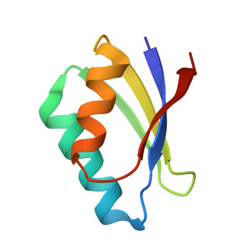
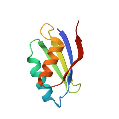
Crystal structure of the adduct HAH1-Cd(II)-MNK1.


Experimental Data Snapshot

  • Method: X-RAY DIFFRACTION
  • Resolution: 1.80 Å
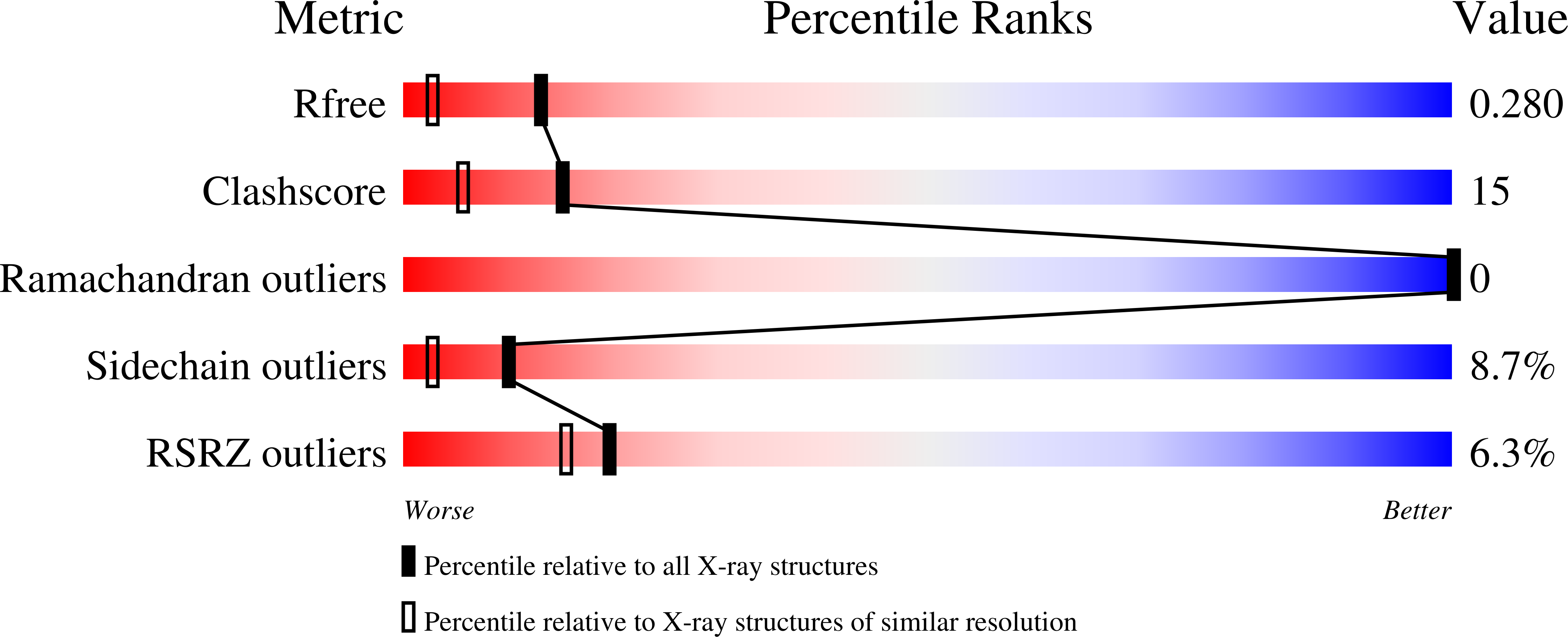
  • R-Value Free: 0.283 
  • R-Value Work: 0.226 
  • R-Value Observed: 0.231 

Starting Model: experimental
View more details

wwPDB Validation   3D Report Full Report


This is version 1.2 of the entry. See complete history


Literature

Copper(I)-mediated protein-protein interactions result from suboptimal interaction surfaces.

Banci, L.Bertini, I.Calderone, V.Della-Malva, N.Felli, I.C.Neri, S.Pavelkova, A.Rosato, A.

(2009) Biochem J 422: 37-42

  • DOI: https://doi.org/10.1042/BJ20090422
  • Primary Citation of Related Structures:  
    3CJK

  • PubMed Abstract: 

    The homoeostasis of metal ions in cells is the result of the contribution of several cellular pathways that involve transient, often weak, protein-protein interactions. Metal transfer typically implies the formation of adducts where the metal itself acts as a bridge between proteins, by co-ordinating residues of both interacting partners. In the present study we address the interaction between the human copper(I)-chaperone HAH1 (human ATX1 homologue) and a metal-binding domain in one of its partners, namely the P-type copper-transporting ATPase, ATP7A (ATPase, Cu+ transporting, alpha polypeptide). The adduct was structurally characterized in solution, in the presence of copper(I), and through X-ray crystallography, upon replacing copper(I) with cadmium(II). Further insight was obtained through molecular modelling techniques and site-directed mutagenesis. It was found that the interaction involves a relatively small interface (less than 1000 A(2), 1 A=0.1 nm) with a low fraction of non-polar atoms. These observations provide a possible explanation for the low affinity of the two apoproteins. It appears that electrostatics is important in selecting which domain of the ATPase is able to form detectable amounts of the metal-mediated adduct with HAH1.


  • Organizational Affiliation

    Magnetic Resonance Center (CERM), University of Florence, Sesto Fiorentino, Italy.


Macromolecules
Find similar proteins by:  (by identity cutoff)  |  3D Structure
Entity ID: 1
MoleculeChains Sequence LengthOrganismDetailsImage
Copper transport protein ATOX168Homo sapiensMutation(s): 0 
Gene Names: ATOX1HAH1
UniProt & NIH Common Fund Data Resources
Find proteins for O00244 (Homo sapiens)
Explore O00244 
Go to UniProtKB:  O00244
PHAROS:  O00244
GTEx:  ENSG00000177556 
Entity Groups  
Sequence Clusters30% Identity50% Identity70% Identity90% Identity95% Identity100% Identity
UniProt GroupO00244
Sequence Annotations
Expand
  • Reference Sequence
Find similar proteins by:  (by identity cutoff)  |  3D Structure
Entity ID: 2
MoleculeChains Sequence LengthOrganismDetailsImage
Copper-transporting ATPase 175Homo sapiensMutation(s): 0 
Gene Names: ATP7AMC1MNK
EC: 3.6.3.4 (PDB Primary Data), 7.2.2.8 (UniProt)
UniProt & NIH Common Fund Data Resources
Find proteins for Q04656 (Homo sapiens)
Explore Q04656 
Go to UniProtKB:  Q04656
PHAROS:  Q04656
GTEx:  ENSG00000165240 
Entity Groups  
Sequence Clusters30% Identity50% Identity70% Identity90% Identity95% Identity100% Identity
UniProt GroupQ04656
Sequence Annotations
Expand
  • Reference Sequence
Small Molecules
Ligands 1 Unique
IDChains Name / Formula / InChI Key2D Diagram3D Interactions
CD
Query on CD

Download Ideal Coordinates CCD File 
C [auth B]CADMIUM ION
Cd
WLZRMCYVCSSEQC-UHFFFAOYSA-N
Experimental Data & Validation

Experimental Data

  • Method: X-RAY DIFFRACTION
  • Resolution: 1.80 Å
  • R-Value Free: 0.283 
  • R-Value Work: 0.226 
  • R-Value Observed: 0.231 
  • Space Group: P 21 21 21
Unit Cell:
Length ( Å )Angle ( ˚ )
a = 47.707α = 90
b = 55.274β = 90
c = 63.241γ = 90
Software Package:
Software NamePurpose
REFMACrefinement
ADSCdata collection
MOSFLMdata reduction
SCALAdata scaling
MOLREPphasing

Structure Validation

View Full Validation Report


Entry History 

Deposition Data

Revision History  (Full details and data files)

  • Version 1.0: 2008-12-30
    Type: Initial release
  • Version 1.1: 2011-07-13
    Changes: Version format compliance
  • Version 1.2: 2023-08-30
    Changes: Data collection, Database references, Derived calculations, Refinement description