Crystal structure of Escherichia coli CheY refined at 1.7-A resolution.
Volz, K., Matsumura, P.(1991) J Biol Chem 266: 15511-15519
- PubMed: 1869568
- DOI: https://doi.org/10.2210/pdb3chy/pdb
- Primary Citation of Related Structures:
3CHY - PubMed Abstract:
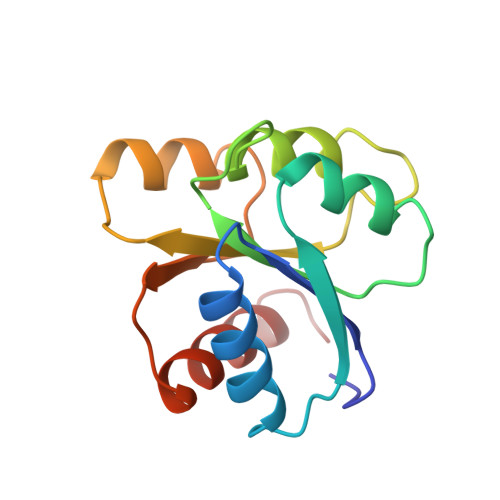
The three-dimensional structure of wild-type CheY from Escherichia coli has been refined by stereochemically restrained least squares minimization to a crystallographic R-factor of 15.1% at 1.7-A resolution. The structure contains 1165 atoms, including all atoms of the protein, 147 water molecules, and three sulfate ions. The final model has root mean square deviations of 0.018 and 0.049 A from idealized bond lengths and angle distances, respectively. Seven amino acid side chains have been modeled in dual conformations. CheY folds as a compact (beta/alpha)5 globular protein, with the phosphorylation region contained in a cavity on one face of the molecule. This active site area is bordered by the carboxyl termini of the three central beta-strands, by alpha 1, and by the loop connecting beta 5 to alpha 5. The Lys-109 side chain of this loop extends into the active site by virtue of its cis peptide bond conformation preceding Pro-110. The epsilon-amino group of Lys-109 is in close bonding contact with the carboxyl group of Asp-57, the residue that is phosphorylated in the activation process of CheY. The details of the hydrogen bonding network in the phosphorylation region indicate that structural rearrangements must accompany the phosphorylation of Asp-57.
Organizational Affiliation:
Department of Microbiology and Immunology, University of Illinois, Chicago 60612.