3BGC

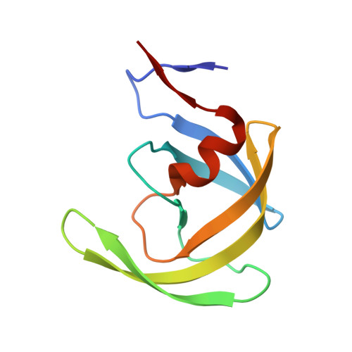
HIV-1 protease in complex with a benzyl decorated oligoamine


Experimental Data Snapshot

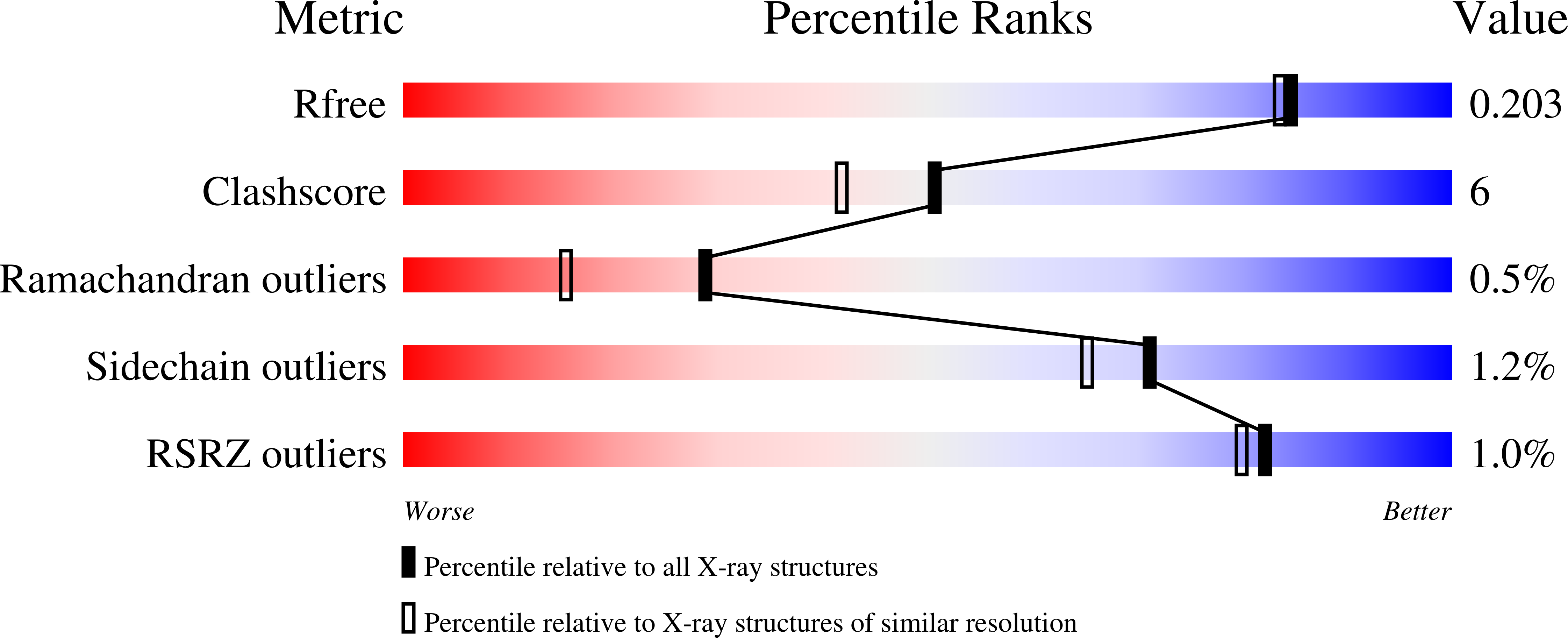
  • Method: X-RAY DIFFRACTION
  • Resolution: 1.80 Å
  • R-Value Free: 0.230 
  • R-Value Work: 0.184 
  • R-Value Observed: 0.185 

Starting Model: experimental
View more details

wwPDB Validation   3D Report Full Report


Ligand Structure Quality Assessment 


This is version 1.2 of the entry. See complete history


Literature

Achiral oligoamines as versatile tool for the development of aspartic protease inhibitors

Blum, A.Sammet, B.Luksch, T.Heine, A.Klebe, G.Diederich, W.E.

(2008) Bioorg Med Chem 16: 8574-8586

  • DOI: https://doi.org/10.1016/j.bmc.2008.08.012
  • Primary Citation of Related Structures:  
    3BGB, 3BGC

  • PubMed Abstract: 

    Due to the important role that aspartic proteases play in many patho-physiological processes, they have intensively been targeted by modern drug development. However, up to now, only for two family members, renin and HIV protease, approved drugs are available. Inhibitor development, mostly guided by mimicking the natural peptide substrates, resulted in very potent inhibitors for several targets, but the pharmacokinetic properties of these compounds were often not optimal. Herein we report a novel approach for lead structure discovery of non-peptidic aspartic protease inhibitors using easily accessible achiral linear oligoamines as starting point. An initial library comprising 11 inhibitors was developed and screened against six selected aspartic proteases. Several hits could be identified, among them selective as well as rather promiscuous inhibitors. The design concept was confirmed by determination of the crystal structure of two derivatives in complex with the HIV-1 protease, and represents a promising basis for the further inhibitor development.


  • Organizational Affiliation

    Institut für Pharmazeutische Chemie, Philipps-Universität Marburg, Marbacher Weg 6, 35032 Marburg, Germany.


Macromolecules
Find similar proteins by:  (by identity cutoff)  |  3D Structure
Entity ID: 1
MoleculeChains Sequence LengthOrganismDetailsImage
Protease
A, B
99N/AMutation(s): 0 
Gene Names: gag-pol
EC: 3.4.23.16
UniProt
Find proteins for P04587 (Human immunodeficiency virus type 1 group M subtype B (isolate BH5))
Explore P04587 
Go to UniProtKB:  P04587
Entity Groups  
Sequence Clusters30% Identity50% Identity70% Identity90% Identity95% Identity100% Identity
UniProt GroupP04587
Sequence Annotations
Expand
  • Reference Sequence
Small Molecules
Binding Affinity Annotations 
IDSourceBinding Affinity
LJH BindingDB:  3BGC Ki: 9600 (nM) from 1 assay(s)
PDBBind:  3BGC Ki: 9600 (nM) from 1 assay(s)
Experimental Data & Validation

Experimental Data

  • Method: X-RAY DIFFRACTION
  • Resolution: 1.80 Å
  • R-Value Free: 0.230 
  • R-Value Work: 0.184 
  • R-Value Observed: 0.185 
  • Space Group: P 21 21 2
Unit Cell:
Length ( Å )Angle ( ˚ )
a = 57.43α = 90
b = 85.724β = 90
c = 46.54γ = 90
Software Package:
Software NamePurpose
SHELXmodel building
SHELXL-97refinement
CrystalCleardata collection
HKL-2000data reduction
HKL-2000data scaling
PHASERphasing

Structure Validation

View Full Validation Report


Ligand Structure Quality Assessment 


Entry History 

Deposition Data

Revision History  (Full details and data files)

  • Version 1.0: 2008-09-02
    Type: Initial release
  • Version 1.1: 2011-07-13
    Changes: Version format compliance
  • Version 1.2: 2023-11-01
    Changes: Data collection, Database references, Derived calculations, Refinement description