2V0X

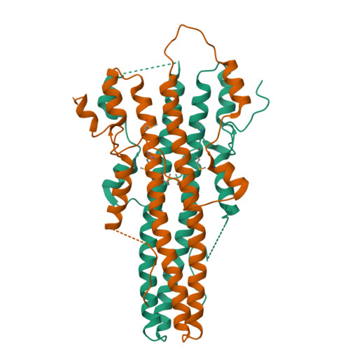
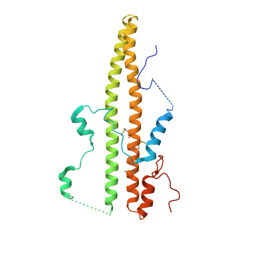
The dimerization domain of LAP2alpha


Experimental Data Snapshot

  • Method: X-RAY DIFFRACTION
  • Resolution: 2.20 Å
  • R-Value Free: 0.257 
  • R-Value Work: 0.213 

wwPDB Validation   3D Report Full Report


This is version 1.5 of the entry. See complete history


Literature

Structural Basis for Dimerization of Lap2Alpha, a Component of the Nuclear Lamina.

Bradley, C.M.Jones, S.Huang, Y.Suzuki, Y.Kvaratskhelia, M.Hickman, A.B.Craigie, R.Dyda, F.

(2007) Structure 15: 643

  • DOI: https://doi.org/10.1016/j.str.2007.04.007
  • Primary Citation of Related Structures:  
    2V0X

  • PubMed Abstract: 

    Lamina-associated polypeptides (LAPs) are important components of the nuclear lamina, the dense network of filaments that supports the nuclear envelope and also extends into the nucleoplasm. The main protein constituents of the nuclear lamina are the constitutively expressed B-type lamins and the developmentally regulated A- and C-type lamins. LAP2alpha is the only non-membrane-associated member of the LAP family. It preferentially binds lamin A/C, has been implicated in cell-cycle regulation and chromatin organization, and has also been found to be a component of retroviral preintegration complexes. As an approach to understanding the role of LAP2alpha in cellular pathways, we have determined the crystal structure of the C-terminal domain of LAP2alpha, residues 459-693. The C-terminal domain is dimeric and possesses an extensive four-stranded, antiparallel coiled coil. The surface involved in binding lamin A/C is proposed based on results from alanine-scanning mutagenesis and a solid-phase overlay binding assay.


  • Organizational Affiliation

    Laboratory of Molecular Biology, National Institute of Diabetes and Digestive and Kidney Diseases, National Institutes of Health, Bethesda, MD 20892, USA.


Macromolecules
Find similar proteins by:  (by identity cutoff)  |  3D Structure
Entity ID: 1
MoleculeChains Sequence LengthOrganismDetailsImage
LAMINA-ASSOCIATED POLYPEPTIDE 2 ISOFORMS ALPHA/ZETA
A, B
235Mus musculusMutation(s): 2 
UniProt
Find proteins for Q61033 (Mus musculus)
Explore Q61033 
Go to UniProtKB:  Q61033
Entity Groups  
Sequence Clusters30% Identity50% Identity70% Identity90% Identity95% Identity100% Identity
UniProt GroupQ61033
Sequence Annotations
Expand
  • Reference Sequence
Small Molecules
Modified Residues  1 Unique
IDChains TypeFormula2D DiagramParent
MSE
Query on MSE
A, B
L-PEPTIDE LINKINGC5 H11 N O2 SeMET
Experimental Data & Validation

Experimental Data

  • Method: X-RAY DIFFRACTION
  • Resolution: 2.20 Å
  • R-Value Free: 0.257 
  • R-Value Work: 0.213 
  • Space Group: P 21 21 21
Unit Cell:
Length ( Å )Angle ( ˚ )
a = 55.084α = 90
b = 55.683β = 90
c = 122.911γ = 90
Software Package:
Software NamePurpose
CNXrefinement
HKL-2000data reduction
HKL-2000data scaling
PHASESphasing

Structure Validation

View Full Validation Report


Entry History 

Deposition Data

Revision History  (Full details and data files)

  • Version 1.0: 2007-06-26
    Type: Initial release
  • Version 1.1: 2011-05-08
    Changes: Version format compliance
  • Version 1.2: 2011-07-13
    Changes: Version format compliance
  • Version 1.3: 2019-03-06
    Changes: Data collection, Derived calculations, Experimental preparation, Other
  • Version 1.4: 2019-05-15
    Changes: Data collection, Experimental preparation
  • Version 1.5: 2024-10-16
    Changes: Data collection, Database references, Structure summary