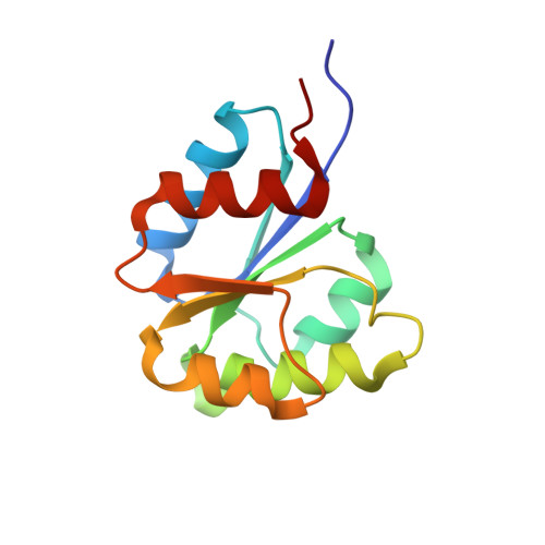
2NT4

Receiver domain from Myxococcus xanthus social motility protein FrzS (H92F mutant)


Experimental Data Snapshot

  • Method: X-RAY DIFFRACTION
  • Resolution: 1.02 Å
  • R-Value Free: 0.154 
  • R-Value Work: 0.123 
  • R-Value Observed: 0.124 

Starting Model: experimental
View more details

wwPDB Validation   3D Report Full Report


This is version 1.4 of the entry. See complete history


Literature

An atypical receiver domain controls the dynamic polar localization of the Myxococcus xanthus social motility protein FrzS.

Fraser, J.S.Merlie, J.P.Echols, N.Weisfield, S.R.Mignot, T.Wemmer, D.E.Zusman, D.R.Alber, T.

(2007) Mol Microbiol 65: 319-332

  • DOI: https://doi.org/10.1111/j.1365-2958.2007.05785.x
  • Primary Citation of Related Structures:  
    2GKG, 2I6F, 2NT3, 2NT4

  • PubMed Abstract: 

    The Myxococcus xanthus FrzS protein transits from pole-to-pole within the cell, accumulating at the pole that defines the direction of movement in social (S) motility. Here we show using atomic-resolution crystallography and NMR that the FrzS receiver domain (RD) displays the conserved switch Tyr102 in an unusual conformation, lacks the conserved Asp phosphorylation site, and fails to bind Mg(2+) or the phosphoryl analogue, Mg(2+) x BeF(3). Mutation of Asp55, closest to the canonical site of RD phosphorylation, showed no motility phenotype in vivo, demonstrating that phosphorylation at this site is not necessary for domain function. In contrast, the Tyr102Ala and His92Phe substitutions on the canonical output face of the FrzS RD abolished S-motility in vivo. Single-cell fluorescence microscopy measurements revealed a striking mislocalization of these mutant FrzS proteins to the trailing cell pole in vivo. The crystal structures of the mutants suggested that the observed conformation of Tyr102 in the wild-type FrzS RD is not sufficient for function. These results support the model that FrzS contains a novel 'pseudo-receiver domain' whose function requires recognition of the RD output face but not Asp phosphorylation.


  • Organizational Affiliation

    Department of Molecular and Cell Biology, University of California, Berkeley, CA 94720-3320, USA.


Macromolecules
Find similar proteins by:  (by identity cutoff)  |  3D Structure
Entity ID: 1
MoleculeChains Sequence LengthOrganismDetailsImage
Response regulator homolog127Myxococcus xanthusMutation(s): 1 
Gene Names: frzS
UniProt
Find proteins for O68522 (Myxococcus xanthus)
Explore O68522 
Go to UniProtKB:  O68522
Entity Groups  
Sequence Clusters30% Identity50% Identity70% Identity90% Identity95% Identity100% Identity
UniProt GroupO68522
Sequence Annotations
Expand
  • Reference Sequence
Small Molecules
Ligands 1 Unique
IDChains Name / Formula / InChI Key2D Diagram3D Interactions
CL
Query on CL

Download Ideal Coordinates CCD File 
B [auth A]CHLORIDE ION
Cl
VEXZGXHMUGYJMC-UHFFFAOYSA-M
Experimental Data & Validation

Experimental Data

  • Method: X-RAY DIFFRACTION
  • Resolution: 1.02 Å
  • R-Value Free: 0.154 
  • R-Value Work: 0.123 
  • R-Value Observed: 0.124 
  • Space Group: P 1 21 1
Unit Cell:
Length ( Å )Angle ( ˚ )
a = 34.883α = 90
b = 37.119β = 102.49
c = 42.104γ = 90
Software Package:
Software NamePurpose
REFMACrefinement
HKL-2000data reduction
HKL-2000data scaling
MOLREPphasing

Structure Validation

View Full Validation Report


Entry History 

Deposition Data

Revision History  (Full details and data files)

  • Version 1.0: 2007-03-13
    Type: Initial release
  • Version 1.1: 2008-05-01
    Changes: Version format compliance
  • Version 1.2: 2011-07-13
    Changes: Version format compliance
  • Version 1.3: 2021-10-20
    Changes: Database references, Derived calculations
  • Version 1.4: 2023-08-30
    Changes: Data collection, Refinement description