2BNP

Lipidic cubic phase grown reaction centre from Rhodobacter sphaeroides, ground state


Experimental Data Snapshot

  • Method: X-RAY DIFFRACTION
  • Resolution: 2.70 Å
  • R-Value Free: 0.249 
  • R-Value Work: 0.210 
  • R-Value Observed: 0.212 

Starting Model: experimental
View more details

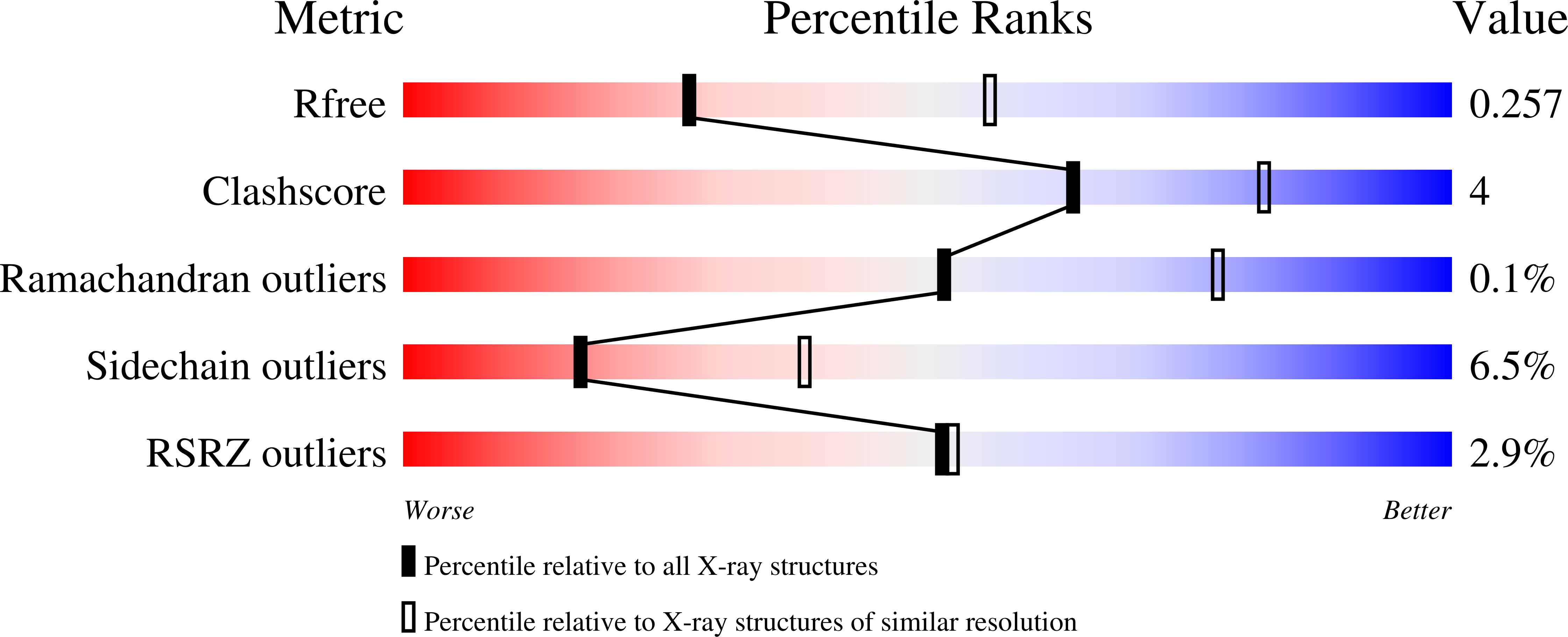
wwPDB Validation   3D Report Full Report


Ligand Structure Quality Assessment 


This is version 1.3 of the entry. See complete history


Literature

Conformational Regulation of Charge Recombination Reactions in a Photosynthetic Bacterial Reaction Centre

Katona, G.Snijder, A.Gourdon, P.Andreasson, U.Hansson, O.Andreasson, L.E.Neutze, R.

(2005) Nat Struct Mol Biol 12: 630

  • DOI: https://doi.org/10.1038/nsmb948
  • Primary Citation of Related Structures:  
    2BNP, 2BNS

  • PubMed Abstract: 

    In bright light the photosynthetic reaction center (RC) of Rhodobacter sphaeroides stabilizes the P(+)(870).Q(-)(A) charge-separated state and thereby minimizes the potentially harmful effects of light saturation. Using X-ray diffraction we report a conformational change that occurs within the cytoplasmic domain of this RC in response to prolonged illumination with bright light. Our observations suggest a novel structural mechanism for the regulation of electron transfer reactions in photosynthesis.


  • Organizational Affiliation

    Chalmers University of Technology, Department of Chemistry and Bioscience, PO Box 462, SE-405 30 Göteborg, Sweden.


Macromolecules
Find similar proteins by:  (by identity cutoff)  |  3D Structure
Entity ID: 1
MoleculeChains Sequence LengthOrganismDetailsImage
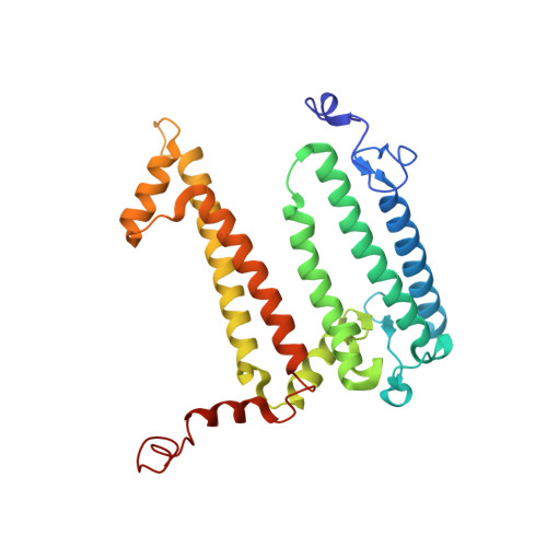
Reaction center protein L chain281Cereibacter sphaeroidesMutation(s): 0 
Membrane Entity: Yes 
UniProt
Find proteins for P0C0Y8 (Cereibacter sphaeroides)
Explore P0C0Y8 
Go to UniProtKB:  P0C0Y8
Entity Groups  
Sequence Clusters30% Identity50% Identity70% Identity90% Identity95% Identity100% Identity
UniProt GroupP0C0Y8
Sequence Annotations
Expand
  • Reference Sequence
Find similar proteins by:  (by identity cutoff)  |  3D Structure
Entity ID: 2
MoleculeChains Sequence LengthOrganismDetailsImage
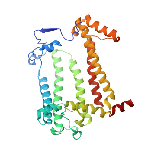
Reaction center protein M chain307Cereibacter sphaeroidesMutation(s): 0 
Membrane Entity: Yes 
UniProt
Find proteins for P0C0Y9 (Cereibacter sphaeroides)
Explore P0C0Y9 
Go to UniProtKB:  P0C0Y9
Entity Groups  
Sequence Clusters30% Identity50% Identity70% Identity90% Identity95% Identity100% Identity
UniProt GroupP0C0Y9
Sequence Annotations
Expand
  • Reference Sequence
Find similar proteins by:  (by identity cutoff)  |  3D Structure
Entity ID: 3
MoleculeChains Sequence LengthOrganismDetailsImage
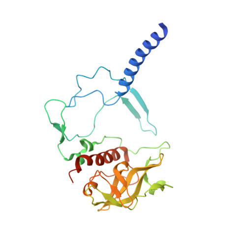
Reaction center protein H chain260Cereibacter sphaeroidesMutation(s): 0 
Membrane Entity: Yes 
UniProt
Find proteins for P0C0Y7 (Cereibacter sphaeroides)
Explore P0C0Y7 
Go to UniProtKB:  P0C0Y7
Entity Groups  
Sequence Clusters30% Identity50% Identity70% Identity90% Identity95% Identity100% Identity
UniProt GroupP0C0Y7
Sequence Annotations
Expand
  • Reference Sequence
Small Molecules
Ligands 7 Unique
IDChains Name / Formula / InChI Key2D Diagram3D Interactions
BCL
Query on BCL

Download Ideal Coordinates CCD File 
D [auth A],
E [auth A],
H [auth B],
I [auth B]
BACTERIOCHLOROPHYLL A
C55 H74 Mg N4 O6
DSJXIQQMORJERS-AGGZHOMASA-M
BPH
Query on BPH

Download Ideal Coordinates CCD File 
F [auth A],
J [auth B]
BACTERIOPHEOPHYTIN A
C55 H76 N4 O6
KWOZSBGNAHVCKG-SZQBJALDSA-N
U10
Query on U10

Download Ideal Coordinates CCD File 
L [auth B]UBIQUINONE-10
C59 H90 O4
ACTIUHUUMQJHFO-UPTCCGCDSA-N
MST
Query on MST

Download Ideal Coordinates CCD File 
G [auth A]2-T-BUTYLAMINO-4-ETHYLAMINO-6-METHYLTHIO-S-TRIAZINE
C10 H19 N5 S
IROINLKCQGIITA-UHFFFAOYSA-N
PO4
Query on PO4

Download Ideal Coordinates CCD File 
N [auth B],
O [auth B]
PHOSPHATE ION
O4 P
NBIIXXVUZAFLBC-UHFFFAOYSA-K
FE2
Query on FE2

Download Ideal Coordinates CCD File 
K [auth B]FE (II) ION
Fe
CWYNVVGOOAEACU-UHFFFAOYSA-N
CL
Query on CL

Download Ideal Coordinates CCD File 
M [auth B]CHLORIDE ION
Cl
VEXZGXHMUGYJMC-UHFFFAOYSA-M
Experimental Data & Validation

Experimental Data

  • Method: X-RAY DIFFRACTION
  • Resolution: 2.70 Å
  • R-Value Free: 0.249 
  • R-Value Work: 0.210 
  • R-Value Observed: 0.212 
  • Space Group: P 42 21 2
Unit Cell:
Length ( Å )Angle ( ˚ )
a = 100.142α = 90
b = 100.142β = 90
c = 238.002γ = 90
Software Package:
Software NamePurpose
REFMACrefinement
MOSFLMdata reduction
SCALAdata scaling

Structure Validation

View Full Validation Report


Ligand Structure Quality Assessment 


Entry History 

Deposition Data

Revision History  (Full details and data files)

  • Version 1.0: 2005-05-27
    Type: Initial release
  • Version 1.1: 2013-03-20
    Changes: Atomic model, Derived calculations, Non-polymer description, Other, Structure summary, Version format compliance
  • Version 1.2: 2019-09-25
    Changes: Advisory, Data collection, Database references, Experimental preparation, Other, Source and taxonomy, Structure summary
  • Version 1.3: 2023-12-13
    Changes: Advisory, Data collection, Database references, Derived calculations, Refinement description