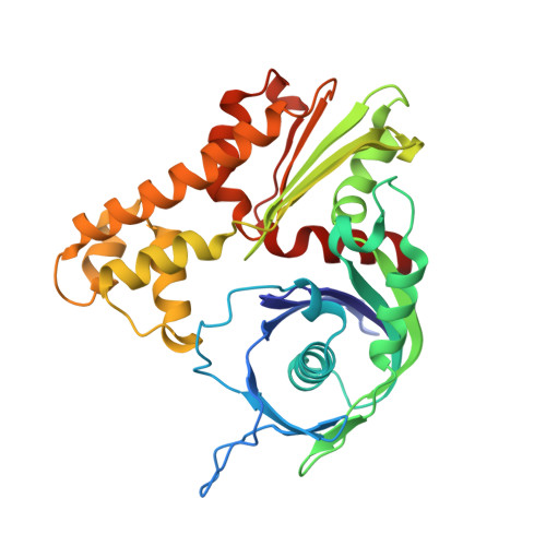
Molecular structure of the ParM polymer and the mechanism leading to its nucleotide-driven dynamic instability
Popp, D., Narita, A., Oda, T., Fujisawa, T., Matsuo, H., Nitanai, Y., Iwasa, M., Maeda, K., Onishi, H., Maeda, Y.(2008) EMBO J 27: 570-579
- PubMed: 18188150
- DOI: https://doi.org/10.1038/sj.emboj.7601978
- Primary Citation of Related Structures:
2ZGY, 2ZGZ, 2ZHC - PubMed Abstract:
ParM is a prokaryotic actin homologue, which ensures even plasmid segregation before bacterial cell division. In vivo, ParM forms a labile filament bundle that is reminiscent of the more complex spindle formed by microtubules partitioning chromosomes in eukaryotic cells. However, little is known about the underlying structural mechanism of DNA segregation by ParM filaments and the accompanying dynamic instability. Our biochemical, TIRF microscopy and high-pressure SAX observations indicate that polymerization and disintegration of ParM filaments is driven by GTP rather than ATP and that ParM acts as a GTP-driven molecular switch similar to a G protein. Image analysis of electron micrographs reveals that the ParM filament is a left-handed helix, opposed to the right-handed actin polymer. Nevertheless, the intersubunit contacts are similar to those of actin. Our atomic model of the ParM-GMPPNP filament, which also fits well to X-ray fibre diffraction patterns from oriented gels, can explain why after nucleotide release, large conformational changes of the protomer lead to a breakage of intra- and interstrand interactions, and thus to the observed disintegration of the ParM filament after DNA segregation.
Organizational Affiliation:
ERATO Actin Filament Dynamics Project, RIKEN Harima Institute, Japan Science and Technology Corporation, Sayo, Hyogo, Japan. dpopp@spring8.or.jp