2ZBD

Crystal Structure of the SR Calcium Pump with Bound Aluminium Fluoride, ADP and Calcium


Experimental Data Snapshot

  • Method: X-RAY DIFFRACTION
  • Resolution: 2.40 Å
  • R-Value Free: 0.271 
  • R-Value Work: 0.246 
  • R-Value Observed: 0.248 

wwPDB Validation   3D Report Full Report


This is version 1.2 of the entry. See complete history


Literature

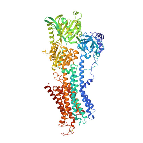
Lumenal gating mechanism revealed in calcium pump crystal structures with phosphate analogues

Toyoshima, C.Nomura, H.Tsuda, T.

(2004) Nature 432: 361-368

  • DOI: https://doi.org/10.1038/nature02981
  • Primary Citation of Related Structures:  
    1WPG, 2ZBD

  • PubMed Abstract: 

    P-type ion transporting ATPases are ATP-powered ion pumps that establish ion concentration gradients across biological membranes. Transfer of bound cations to the lumenal or extracellular side occurs while the ATPase is phosphorylated. Here we report at 2.3 A resolution the structure of the calcium-ATPase of skeletal muscle sarcoplasmic reticulum, a representative P-type ATPase that is crystallized in the absence of Ca2+ but in the presence of magnesium fluoride, a stable phosphate analogue. This and other crystal structures determined previously provide atomic models for all four principal states in the reaction cycle. These structures show that the three cytoplasmic domains rearrange to move six out of ten transmembrane helices, thereby changing the affinity of the Ca2+-binding sites and the gating of the ion pathway. Release of ADP triggers the opening of the lumenal gate and release of phosphate its closure, effected mainly through movement of the A-domain, the actuator of transmembrane gates.


  • Organizational Affiliation

    Institute of Molecular and Cellular Biosciences, The University of Tokyo, Bunkyo-ku, Tokyo 113-0032, Japan. ct@iam.u-tokyo.ac.jp


Macromolecules
Find similar proteins by:  (by identity cutoff)  |  3D Structure
Entity ID: 1
MoleculeChains Sequence LengthOrganismDetailsImage
Sarcoplasmic/endoplasmic reticulum calcium ATPase 1995Oryctolagus cuniculusMutation(s): 0 
EC: 3.6.3.8
Membrane Entity: Yes 
UniProt
Find proteins for P04191 (Oryctolagus cuniculus)
Explore P04191 
Go to UniProtKB:  P04191
Entity Groups  
Sequence Clusters30% Identity50% Identity70% Identity90% Identity95% Identity100% Identity
UniProt GroupP04191
Sequence Annotations
Expand
  • Reference Sequence
Small Molecules
Ligands 5 Unique
IDChains Name / Formula / InChI Key2D Diagram3D Interactions
PC1
Query on PC1

Download Ideal Coordinates CCD File 
H [auth A],
I [auth A]
1,2-DIACYL-SN-GLYCERO-3-PHOSPHOCHOLINE
C44 H88 N O8 P
NRJAVPSFFCBXDT-HUESYALOSA-N
ADP
Query on ADP

Download Ideal Coordinates CCD File 
G [auth A]ADENOSINE-5'-DIPHOSPHATE
C10 H15 N5 O10 P2
XTWYTFMLZFPYCI-KQYNXXCUSA-N
ALF
Query on ALF

Download Ideal Coordinates CCD File 
E [auth A]TETRAFLUOROALUMINATE ION
Al F4
UYOMQIYKOOHAMK-UHFFFAOYSA-J
CA
Query on CA

Download Ideal Coordinates CCD File 
B [auth A],
C [auth A]
CALCIUM ION
Ca
BHPQYMZQTOCNFJ-UHFFFAOYSA-N
MG
Query on MG

Download Ideal Coordinates CCD File 
D [auth A],
F [auth A]
MAGNESIUM ION
Mg
JLVVSXFLKOJNIY-UHFFFAOYSA-N
Experimental Data & Validation

Experimental Data

  • Method: X-RAY DIFFRACTION
  • Resolution: 2.40 Å
  • R-Value Free: 0.271 
  • R-Value Work: 0.246 
  • R-Value Observed: 0.248 
  • Space Group: C 1 2 1
Unit Cell:
Length ( Å )Angle ( ˚ )
a = 162.9α = 90
b = 75.1β = 109.3
c = 152.1γ = 90
Software Package:
Software NamePurpose
CNSrefinement
REFMACrefinement
DENZOdata reduction
SCALEPACKdata scaling
CNSphasing

Structure Validation

View Full Validation Report


Entry History 

Deposition Data

Revision History  (Full details and data files)

  • Version 1.0: 2007-11-20
    Type: Initial release
  • Version 1.1: 2011-07-13
    Changes: Version format compliance
  • Version 1.2: 2023-11-01
    Changes: Data collection, Database references, Derived calculations, Refinement description