2YFY

SERCA in the HnE2 State Complexed With Debutanoyl Thapsigargin


Experimental Data Snapshot

  • Method: X-RAY DIFFRACTION
  • Resolution: 3.10 Å
  • R-Value Free: 0.278 
  • R-Value Work: 0.239 
  • R-Value Observed: 0.240 

wwPDB Validation   3D Report Full Report


Ligand Structure Quality Assessment 


This is version 1.1 of the entry. See complete history


Literature

Mutual Adaptation of a Membrane Protein and its Lipid Bilayer During Conformational Changes.

Sonntag, Y.Musgaard, M.Olesen, C.Schiott, B.Moller, J.V.Nissen, P.Thogersen, L.

(2011) Nat Commun 2: 304

  • DOI: https://doi.org/10.1038/ncomms1307
  • Primary Citation of Related Structures:  
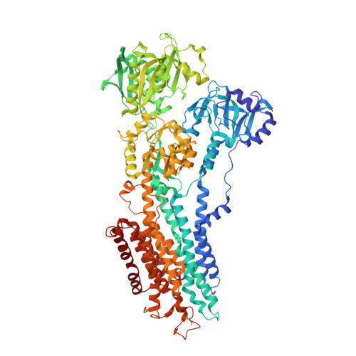
    2YFY

  • PubMed Abstract: 

    The structural elucidation of membrane proteins continues to gather pace, but we know little about their molecular interactions with the lipid environment or how they interact with the surrounding bilayer. Here, with the aid of low-resolution X-ray crystallography, we present direct structural information on membrane interfaces as delineated by lipid phosphate groups surrounding the sarco(endo)plasmic reticulum Ca(2+)-ATPase (SERCA) in its phosphorylated and dephosphorylated Ca(2+)-free forms. The protein-lipid interactions are further analysed using molecular dynamics simulations. We find that SERCA adapts to membranes of different hydrophobic thicknesses by inducing local deformations in the lipid bilayers and by undergoing small rearrangements of the amino-acid side chains and helix tilts. These mutually adaptive interactions allow smooth transitions through large conformational changes associated with the transport cycle of SERCA, a strategy that may be of general nature for many membrane proteins.


  • Organizational Affiliation

    Centre for Membrane Pumps in Cells and Disease - PUMPKIN, Danish National Research Foundation, DK-8000 Aarhus C, Denmark.


Macromolecules
Find similar proteins by:  (by identity cutoff)  |  3D Structure
Entity ID: 1
MoleculeChains Sequence LengthOrganismDetailsImage
SARCOPLASMIC/ENDOPLASMIC RETICULUM CALCIUM ATPASE 1994Oryctolagus cuniculusMutation(s): 0 
EC: 3.6.3.8
Membrane Entity: Yes 
UniProt
Find proteins for P04191 (Oryctolagus cuniculus)
Explore P04191 
Go to UniProtKB:  P04191
Entity Groups  
Sequence Clusters30% Identity50% Identity70% Identity90% Identity95% Identity100% Identity
UniProt GroupP04191
Sequence Annotations
Expand
  • Reference Sequence
Experimental Data & Validation

Experimental Data

  • Method: X-RAY DIFFRACTION
  • Resolution: 3.10 Å
  • R-Value Free: 0.278 
  • R-Value Work: 0.239 
  • R-Value Observed: 0.240 
  • Space Group: P 41 21 2
Unit Cell:
Length ( Å )Angle ( ˚ )
a = 71.36α = 90
b = 71.36β = 90
c = 587.91γ = 90
Software Package:
Software NamePurpose
PHENIXrefinement
XDSdata reduction
XSCALEdata scaling
PHASERphasing

Structure Validation

View Full Validation Report


Ligand Structure Quality Assessment 


Entry History 

Deposition Data

Revision History  (Full details and data files)

  • Version 1.0: 2011-06-29
    Type: Initial release
  • Version 1.1: 2011-07-13
    Changes: Version format compliance