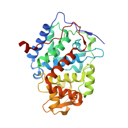
2Y5A

Cytochrome c peroxidase (CCP) W191G bound to 3-aminopyridine


Experimental Data Snapshot

  • Method: X-RAY DIFFRACTION
  • Resolution: 1.25 Å
  • R-Value Free: 0.164 
  • R-Value Work: 0.150 
  • R-Value Observed: 0.150 

wwPDB Validation   3D Report Full Report


Ligand Structure Quality Assessment 


This is version 1.2 of the entry. See complete history


Literature

Probing the Dynamic Nature of Water Molecules and Their Influences on Ligand Binding in a Model Binding Site.

Cappel, D.Wahlstrom, R.Brenk, R.Sotriffer, C.A.

(2011) J Chem Inf Model 51: 2581

  • DOI: https://doi.org/10.1021/ci200052j
  • Primary Citation of Related Structures:  
    2Y5A

  • PubMed Abstract: 

    The model binding site of the cytochrome c peroxidase (CCP) W191G mutant is used to investigate the structural and dynamic properties of the water network at the buried cavity using computational methods supported by crystallographic analysis. In particular, the differences of the hydration pattern between the uncomplexed state and various complexed forms are analyzed as well as the differences between five complexes of CCP W191G with structurally closely related ligands. The ability of docking programs to correctly handle the water molecules in these systems is studied in detail. It is found that fully automated prediction of water replacement or retention upon docking works well if some additional preselection is carried out but not necessarily if the entire water network in the cavity is used as input. On the other hand, molecular interaction fields for water calculated from static crystal structures and hydration density maps obtained from molecular dynamics simulations agree very well with crystallographically observed water positions. For one complex, the docking and MD results sensitively depend on the quality of the starting structure, and agreement is obtained only after redetermination of the crystal structure and refinement at higher resolution.


  • Organizational Affiliation

    Institute of Pharmacy and Food Chemistry, Julius-Maximilians University Würzburg, Am Hubland, D-97074 Würzburg, Germany.


Macromolecules
Find similar proteins by:  (by identity cutoff)  |  3D Structure
Entity ID: 1
MoleculeChains Sequence LengthOrganismDetailsImage
CYTOCHROME C PEROXIDASE\, MITOCHONDRIAL294Saccharomyces cerevisiaeMutation(s): 1 
EC: 1.11.1.5
UniProt
Find proteins for P00431 (Saccharomyces cerevisiae (strain ATCC 204508 / S288c))
Explore P00431 
Go to UniProtKB:  P00431
Entity Groups  
Sequence Clusters30% Identity50% Identity70% Identity90% Identity95% Identity100% Identity
UniProt GroupP00431
Sequence Annotations
Expand
  • Reference Sequence
Small Molecules
Ligands 2 Unique
IDChains Name / Formula / InChI Key2D Diagram3D Interactions
HEM
Query on HEM

Download Ideal Coordinates CCD File 
B [auth A]PROTOPORPHYRIN IX CONTAINING FE
C34 H32 Fe N4 O4
KABFMIBPWCXCRK-RGGAHWMASA-L
3AP
Query on 3AP

Download Ideal Coordinates CCD File 
C [auth A]3-AMINOPYRIDINE
C5 H7 N2
CUYKNJBYIJFRCU-UHFFFAOYSA-O
Binding Affinity Annotations 
IDSourceBinding Affinity
3AP Binding MOAD:  2Y5A Kd: 5.50e+4 (nM) from 1 assay(s)
Experimental Data & Validation

Experimental Data

  • Method: X-RAY DIFFRACTION
  • Resolution: 1.25 Å
  • R-Value Free: 0.164 
  • R-Value Work: 0.150 
  • R-Value Observed: 0.150 
  • Space Group: P 21 21 21
Unit Cell:
Length ( Å )Angle ( ˚ )
a = 50.853α = 90
b = 75.362β = 90
c = 106.884γ = 90
Software Package:
Software NamePurpose
REFMACrefinement
DENZOdata reduction
SCALEPACKdata scaling
MOLREPphasing

Structure Validation

View Full Validation Report


Ligand Structure Quality Assessment 


Entry History 

Deposition Data

Revision History  (Full details and data files)

  • Version 1.0: 2011-10-12
    Type: Initial release
  • Version 1.1: 2011-11-02
    Changes: Database references
  • Version 1.2: 2023-12-20
    Changes: Data collection, Database references, Derived calculations, Other, Refinement description