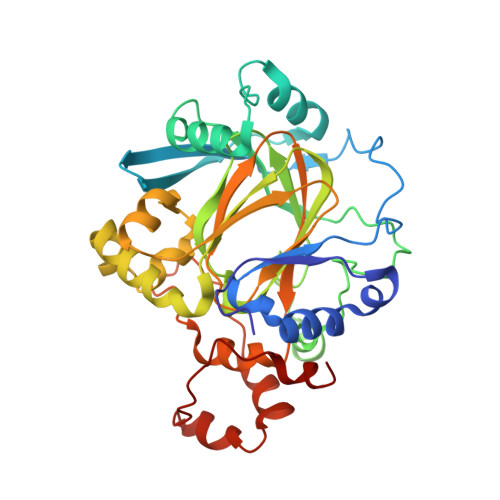
2WWJ

STRUCTURE OF JMJD2A COMPLEXED WITH INHIBITOR 10A


Experimental Data Snapshot

  • Method: X-RAY DIFFRACTION
  • Resolution: 2.60 Å
  • R-Value Free: 0.248 
  • R-Value Work: 0.178 
  • R-Value Observed: 0.181 

Starting Model: experimental
View more details

wwPDB Validation   3D Report Full Report


Ligand Structure Quality Assessment 


This is version 1.4 of the entry. See complete history


Literature

Selective inhibitors of the JMJD2 histone demethylases: combined nondenaturing mass spectrometric screening and crystallographic approaches.

Rose, N.R.Woon, E.C.Kingham, G.L.King, O.N.Mecinovic, J.Clifton, I.J.Ng, S.S.Talib-Hardy, J.Oppermann, U.McDonough, M.A.Schofield, C.J.

(2010) J Med Chem 53: 1810-1818

  • DOI: https://doi.org/10.1021/jm901680b
  • Primary Citation of Related Structures:  
    2WWJ

  • PubMed Abstract: 

    Ferrous ion and 2-oxoglutarate (2OG) oxygenases catalyze the demethylation of N(epsilon)-methylated lysine residues in histones. Here we report studies on the inhibition of the JMJD2 subfamily of histone demethylases, employing binding analyses by nondenaturing mass spectrometry (MS), dynamic combinatorial chemistry coupled to MS, turnover assays, and crystallography. The results of initial binding and inhibition assays directed the production and analysis of a set of N-oxalyl-d-tyrosine derivatives to explore the extent of a subpocket at the JMJD2 active site. Some of the inhibitors were shown to be selective for JMJD2 over the hypoxia-inducible factor prolyl hydroxylase PHD2. A crystal structure of JMJD2A in complex with one of the potent inhibitors was obtained; modeling other inhibitors based on this structure predicts interactions that enable improved inhibition for some compounds.


  • Organizational Affiliation

    The Department of Chemistry and the Oxford Centre for Integrative Systems Biology, Chemistry Research Laboratory, University of Oxford, 12 Mansfield Road, Oxford, OX1 3TA, United Kingdom.


Macromolecules
Find similar proteins by:  (by identity cutoff)  |  3D Structure
Entity ID: 1
MoleculeChains Sequence LengthOrganismDetailsImage
LYSINE-SPECIFIC DEMETHYLASE 4A
A, B
348Homo sapiensMutation(s): 0 
EC: 1.14.11.27 (PDB Primary Data), 1.14.11.66 (UniProt), 1.14.11.69 (UniProt)
UniProt & NIH Common Fund Data Resources
Find proteins for O75164 (Homo sapiens)
Explore O75164 
Go to UniProtKB:  O75164
PHAROS:  O75164
GTEx:  ENSG00000066135 
Entity Groups  
Sequence Clusters30% Identity50% Identity70% Identity90% Identity95% Identity100% Identity
UniProt GroupO75164
Sequence Annotations
Expand
  • Reference Sequence
Small Molecules
Binding Affinity Annotations 
IDSourceBinding Affinity
Y28 PDBBind:  2WWJ IC50: 3.30e+4 (nM) from 1 assay(s)
BindingDB:  2WWJ IC50: 3.30e+4 (nM) from 1 assay(s)
Experimental Data & Validation

Experimental Data

  • Method: X-RAY DIFFRACTION
  • Resolution: 2.60 Å
  • R-Value Free: 0.248 
  • R-Value Work: 0.178 
  • R-Value Observed: 0.181 
  • Space Group: P 21 21 2
Unit Cell:
Length ( Å )Angle ( ˚ )
a = 101.33α = 90
b = 148.97β = 90
c = 57.11γ = 90
Software Package:
Software NamePurpose
REFMACrefinement
MOSFLMdata reduction
SCALAdata scaling
REFMACphasing

Structure Validation

View Full Validation Report


Ligand Structure Quality Assessment 


Entry History 

Deposition Data

Revision History  (Full details and data files)

  • Version 1.0: 2010-03-09
    Type: Initial release
  • Version 1.1: 2011-05-08
    Changes: Version format compliance
  • Version 1.2: 2011-07-13
    Changes: Version format compliance
  • Version 1.3: 2018-03-07
    Changes: Database references, Source and taxonomy, Structure summary
  • Version 1.4: 2023-12-20
    Changes: Data collection, Database references, Derived calculations, Other, Refinement description