2VUK

Structure of the p53 core domain mutant Y220C bound to the stabilizing small-molecule drug PhiKan083


Experimental Data Snapshot

  • Method: X-RAY DIFFRACTION
  • Resolution: 1.50 Å
  • R-Value Free: 0.208 
  • R-Value Work: 0.186 
  • R-Value Observed: 0.186 

wwPDB Validation   3D Report Full Report


Ligand Structure Quality Assessment 


This is version 1.4 of the entry. See complete history


Literature

Targeted Rescue of a Destabilized Mutant of P53 by an in Silico Screened Drug.

Boeckler, F.M.Joerger, A.C.Jaggi, G.Rutherford, T.J.Veprintsev, D.B.Fersht, A.R.

(2008) Proc Natl Acad Sci U S A 105: 10360

  • DOI: https://doi.org/10.1073/pnas.0805326105
  • Primary Citation of Related Structures:  
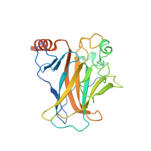
    2VUK

  • PubMed Abstract: 

    The tumor suppressor p53 is mutationally inactivated in approximately 50% of human cancers. Approximately one-third of the mutations lower the melting temperature of the protein, leading to its rapid denaturation. Small molecules that bind to those mutants and stabilize them could be effective anticancer drugs. The mutation Y220C, which occurs in approximately 75,000 new cancer cases per annum, creates a surface cavity that destabilizes the protein by 4 kcal/mol, at a site that is not functional. We have designed a series of binding molecules from an in silico analysis of the crystal structure using virtual screening and rational drug design. One of them, a carbazole derivative (PhiKan083), binds to the cavity with a dissociation constant of approximately 150 muM. It raises the melting temperature of the mutant and slows down its rate of denaturation. We have solved the crystal structure of the protein-PhiKan083 complex at 1.5-A resolution. The structure implicates key interactions between the protein and ligand and conformational changes that occur on binding, which will provide a basis for lead optimization. The Y220C mutant is an excellent "druggable" target for developing and testing novel anticancer drugs based on protein stabilization. We point out some general principles in relationships between binding constants, raising of melting temperatures, and increase of protein half-lives by stabilizing ligands.


  • Organizational Affiliation

    Centre for Protein Engineering, Medical Research Council Centre, Hills Road, Cambridge CB2 0QH, United Kingdom.


Macromolecules
Find similar proteins by:  (by identity cutoff)  |  3D Structure
Entity ID: 1
MoleculeChains Sequence LengthOrganismDetailsImage
CELLULAR TUMOR ANTIGEN P53
A, B
219Homo sapiensMutation(s): 5 
UniProt & NIH Common Fund Data Resources
Find proteins for P04637 (Homo sapiens)
Explore P04637 
Go to UniProtKB:  P04637
PHAROS:  P04637
GTEx:  ENSG00000141510 
Entity Groups  
Sequence Clusters30% Identity50% Identity70% Identity90% Identity95% Identity100% Identity
UniProt GroupP04637
Sequence Annotations
Expand
  • Reference Sequence
Small Molecules
Binding Affinity Annotations 
IDSourceBinding Affinity
P83 Binding MOAD:  2VUK Kd: 1.25e+5 (nM) from 1 assay(s)
PDBBind:  2VUK Kd: 1.25e+5 (nM) from 1 assay(s)
BindingDB:  2VUK Kd: 1.40e+5 (nM) from 1 assay(s)
Experimental Data & Validation

Experimental Data

  • Method: X-RAY DIFFRACTION
  • Resolution: 1.50 Å
  • R-Value Free: 0.208 
  • R-Value Work: 0.186 
  • R-Value Observed: 0.186 
  • Space Group: P 21 21 21
Unit Cell:
Length ( Å )Angle ( ˚ )
a = 65.093α = 90
b = 71.234β = 90
c = 105.207γ = 90
Software Package:
Software NamePurpose
CNSrefinement
CNSphasing

Structure Validation

View Full Validation Report


Ligand Structure Quality Assessment 


Entry History 

Deposition Data

Revision History  (Full details and data files)

  • Version 1.0: 2008-07-22
    Type: Initial release
  • Version 1.1: 2011-05-08
    Changes: Version format compliance
  • Version 1.2: 2011-07-13
    Changes: Version format compliance
  • Version 1.3: 2019-05-08
    Changes: Data collection, Experimental preparation, Other
  • Version 1.4: 2023-12-13
    Changes: Data collection, Database references, Derived calculations, Other, Refinement description