2VAY

Calmodulin complexed with CaV1.1 IQ peptide


Experimental Data Snapshot

  • Method: X-RAY DIFFRACTION
  • Resolution: 1.94 Å
  • R-Value Free: 0.272 
  • R-Value Work: 0.218 
  • R-Value Observed: 0.218 

wwPDB Validation   3D Report Full Report


This is version 1.4 of the entry. See complete history


Literature

Determinants in Cav1 Channels that Regulate the Ca2+ Sensitivity of Bound Calmodulin.

Halling, D.B.Georgiou, D.K.Black, D.J.Yang, G.Fallon, J.L.Quiocho, F.A.Pedersen, S.E.Hamilton, S.L.

(2009) J Biol Chem 284: 20041

  • DOI: https://doi.org/10.1074/jbc.M109.013326
  • Primary Citation of Related Structures:  
    2VAY

  • PubMed Abstract: 

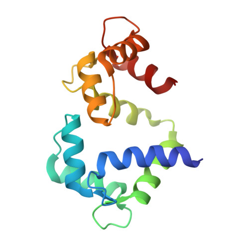
    Calmodulin binds to IQ motifs in the alpha(1) subunit of Ca(V)1.1 and Ca(V)1.2, but the affinities of calmodulin for the motif and for Ca(2+) are higher when bound to Ca(V)1.2 IQ. The Ca(V)1.1 IQ and Ca(V)1.2 IQ sequences differ by four amino acids. We determined the structure of calmodulin bound to Ca(V)1.1 IQ and compared it with that of calmodulin bound to Ca(V)1.2 IQ. Four methionines in Ca(2+)-calmodulin form a hydrophobic binding pocket for the peptide, but only one of the four nonconserved amino acids (His-1532 of Ca(V)1.1 and Tyr-1675 of Ca(V)1.2) contacts this calmodulin pocket. However, Tyr-1675 in Ca(V)1.2 contributes only modestly to the higher affinity of this peptide for calmodulin; the other three amino acids in Ca(V)1.2 contribute significantly to the difference in the Ca(2+) affinity of the bound calmodulin despite having no direct contact with calmodulin. Those residues appear to allow an interaction with calmodulin with one lobe Ca(2+)-bound and one lobe Ca(2+)-free. Our data also provide evidence for lobe-lobe interactions in calmodulin bound to Ca(V)1.2.


  • Organizational Affiliation

    Department of Molecular Physiology and Biophysics, Baylor College of Medicine, Houston, Texas 77030, USA.


Macromolecules
Find similar proteins by:  (by identity cutoff)  |  3D Structure
Entity ID: 1
MoleculeChains Sequence LengthOrganismDetailsImage
CALMODULIN146Homo sapiensMutation(s): 0 
UniProt & NIH Common Fund Data Resources
Find proteins for P0DP23 (Homo sapiens)
Explore P0DP23 
Go to UniProtKB:  P0DP23
PHAROS:  P0DP23
GTEx:  ENSG00000198668 
Entity Groups  
Sequence Clusters30% Identity50% Identity70% Identity90% Identity95% Identity100% Identity
UniProt GroupP0DP23
Sequence Annotations
Expand
  • Reference Sequence

Find similar proteins by:  Sequence   |   3D Structure  

Entity ID: 2
MoleculeChains Sequence LengthOrganismDetailsImage
VOLTAGE-DEPENDENT L-TYPE CALCIUM CHANNEL SUBUNIT ALPHA-1S21Homo sapiensMutation(s): 0 
UniProt & NIH Common Fund Data Resources
Find proteins for Q13698 (Homo sapiens)
Explore Q13698 
Go to UniProtKB:  Q13698
PHAROS:  Q13698
GTEx:  ENSG00000081248 
Entity Groups  
Sequence Clusters30% Identity50% Identity70% Identity90% Identity95% Identity100% Identity
UniProt GroupQ13698
Sequence Annotations
Expand
  • Reference Sequence
Experimental Data & Validation

Experimental Data

  • Method: X-RAY DIFFRACTION
  • Resolution: 1.94 Å
  • R-Value Free: 0.272 
  • R-Value Work: 0.218 
  • R-Value Observed: 0.218 
  • Space Group: C 1 2 1
Unit Cell:
Length ( Å )Angle ( ˚ )
a = 84.92α = 90
b = 34.67β = 113.69
c = 62.98γ = 90
Software Package:
Software NamePurpose
CNSrefinement
HKL-2000data reduction
SCALEPACKdata scaling
CNSphasing

Structure Validation

View Full Validation Report


Entry History 

Deposition Data

Revision History  (Full details and data files)

  • Version 1.0: 2008-09-16
    Type: Initial release
  • Version 1.1: 2011-05-08
    Changes: Version format compliance
  • Version 1.2: 2011-07-13
    Changes: Version format compliance
  • Version 1.3: 2019-05-08
    Changes: Data collection, Experimental preparation, Other
  • Version 1.4: 2023-12-13
    Changes: Data collection, Database references, Derived calculations, Other, Refinement description